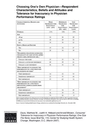
IB SEHS Chapter 1
IB SEHS Chapter 1. Bones. Four Main Types of Bones. Long Bones – usually have a long cylindrical shaft and are enlarged at both ends. They can be large or small but the length is always greater than the width. Long bones are the most important bones for movement.


IB SEHS Chapter 1
E N D
Presentation Transcript
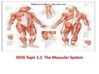
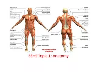
IB SEHSChapter 1 Bones
Four Main Types of Bones • Long Bones – usually have a long cylindrical shaft and are enlarged at both ends. They can be large or small but the length is always greater than the width. Long bones are the most important bones for movement. Long bones include: femur, clavicle, metatarsals
Four Main Types of Bones • Short bones – are small and cube-shaped and they usually articulate with more than one other bone. Short bones include: carpals of the hand, and tarsals of the foot.
Four Main Types of Bones • Flat bones – usually have curved surfaces and vary from being quite thick to very thin. These bones provide protection and the broad surfaces also provide a large area for muscle attachment. Flat bones include: sternum, scapula, ribs, pelvis
Four Main Types of Bones • Irregular bones – have specialized shapes and functions. Irregular bones include: vertebrae, sacrum, coccyx
Another Type of Bone Sesamoid bones – are another type of bone found in the body. These are short bones embedded in tendons where large amounts of pressure develop.
Structure of the Bone *Bones contain a matrix of protein (collagen)fibers along with water and mineral salts. *When the mineral salts accumulate in-between and around the collagen fibers, they crystallize and tissue hardens. *Collagen fibers provide high tensile strength, which allows the bone to resist being stretched or torn apart.
Structure of the Bone The structure of a bone can be described by examining a long bone (see figure 1.6 in your notes). Diaphysis – is the shaft or the mid-section of the long bone. It is made up of compact bone or hard bone. Compact bone is relatively solid and dense, it has few spaces. Compact bone is also found in the outer layer of most other types of bones. It is important for protection, support, and resists the stress of weight placed on long bones.
Structure of the Bone Proximal and Distal Epiphysis (Student check in: What do these words mean?) describe the ends of the bone. They are made up of cancellous or spongy bone. Cancellous bone has an irregular laticework structure where there are many spaces. Cancellous one is also found in short, flat and irregrular bones. Red bone marrow is stored in the cancellous bone and blood cell production occurs here.
Structure of the Bone Articular cartilage - covers the ends of the bones where they articulate (or meet) other bones to form joints. The main functions of the cartilage are to reduce friction between bones an absorb shock. The area not cover by articular cartilage is covered by a thin, shiny white membrane called the periosteum. The periosteum forms the outer lining of bone and is important for bone growth, repair, nutrition and attachment of ligaments and tendons.
Structure of the Bone Medullary (marrow) cavity – is the space within the diaphysis where yellow bone marrow is stored. There is a small opening in the diaphysis called the nutrient foramen. Blood vessels pass through the nutrient foramen and enter the medullary cavity to provide the bone marrow an compact bone with blood and nutrients. Where else did we see a foramen in the body?
In Class Research • Pick a partner to work with. Make your choice wisely. • Using your cell phone (please see me if you do not have a cell phone or internet access on your phone), answer the following questions: a. Identify three areas of the body where sesamoid bones are found. b. What is the largest sesamoid bone in the body? c. It is embedded in the tendon of which muscle? d. What is its main function?
In Class Research Test yourself. Use the skeleton or a picture of a skeleton to help you with these tasks: • Identify the bones of the axial and the appendicular skeleton. • Indicate what type of bone each one is. 3. Most bones are not entirely smooth and have rough areas and prominent landmarks where muscles, tendons and ligaments often attach. See if you can feel the prominent bone landmarks on your arms, legs, trunk. Can you identify which bones these are?