Download

1 / 91
910 likes | 933 Vues
Learn about the monitoring of sleep and wake parameters in congestive heart failure patients, including the diagnosis and types of sleep-related breathing disorders such as obstructive sleep apnea and central sleep apnea syndrome. Recognize the symptoms and signs of these disorders through various scoring models and maneuvers.

E N D
Sleep Related Breathing Disorders In Congestive Heart Failure BYAHMAD YOUNESPROFESSOR OF THORACIC MEDICINE Mansoura Faculty of Medicine
Monitoring of sleep and wake The standard parameters used to record sleep and wake are electroencephalography (EEG), electro-oculography (EOG), electromyography (EMG), airflow measurement, respiratory effort measurement, electrocardiography (ECG), oxygen saturation, snoring monitor, and sleep position evaluation. All these parameters are recorded in polysomnography which is the gold standard for diagnosis of Sleep disordered breathing .
Apnea: is defined as the drop in peak airflow by >90% of baseline for 10 seconds or longer and at least 90% of the event duration meet the amplitude reduction. An obstructive apnea occurs when airflow is absent or nearly absent, but ventilatory effort persists. It is caused by complete, or near complete, upper airway obstruction
A central apnea occurs when both airflow and ventilatory effort are absent.
During a mixed apnea, there is an interval during which there is no respiratory effort (ie, central apnea pattern) and an interval during which there are obstructed respiratory efforts .
Hypopnea • Hypopnea be scored when all of the following criteria are met: 1- Airflow decreases at least 30 percent from baseline 2-There is diminished airflow lasting at least 10 seconds 3- at least 3 percent oxyhemoglobin desaturation . • Apnea-hypopnea index (AHI) is the total number of apneas and hypopneas per hour of sleep. • Respiratory effort related arousal (RERA) is an event characterized by increasing respiratory effort for 10 seconds or longer leading to an arousal from sleep but does not fulfill the criteria for a hypopnea or apnea • The respiratory disturbance index (RDI) is defined as the number of obstructive apneas, hypopneas, and respiratory event–related arousals (RERAs) per hour.
Types of SLEEP RELATED BREATHING DISORDES 1- Obstructive sleep apnea syndrome (OSA) in adults is defined as either • More than 15 apneas, hypopneas, per hour of sleep ( AHI >15 events/hr) in an asymptomatic patient OR • More than 5 apneas, hypopneas, per hour of sleep (AHI >5 events per hour) in a patient with symptoms (eg, sleepiness, fatigue and inattention) or signs of disturbed sleep (snoring, restless sleep, and respiratory pauses).
2- Central sleep apnea syndrome can defined as: a. Study showing AHI > 5 events/hr. and b. CentralAHI > 50% of the total AHI, and c. Central apneas or hypopneas >=5/hr., and d. Symptoms of either excessive sleepiness or disrupted sleep. 3- Sleep Hypoventilation Syndrome if either of the below occur:a-There is an increase in the arterial PaCO2 to a value > 55 mm Hg for ≥ 10 minutes. b. There is ≥ 10 mm Hg increase in PaCO2 during sleep (in comparison to an awake supine value) to a value exceeding 50 mm Hg for ≥ 10 minutes.
In the Mallampati maneuver, patients are instructed not to emit sounds but to open the mouth as wide as possible and protrude the tongue as far as possible. In the modified Mallampati, the patient is instructed to open the mouth as wide as possible without emitting sounds.
Symptoms of central sleep apnea syndrome • Asymptomatic • Excessive sleepiness • Insomnia (repeated nocturnal awakenings) • Nocturnal sensation of dyspnea • Morning headaches • Inattention • Poor concentration
Different forms of CSAS (1) Primary Central Sleep Apnea (2) Central Sleep Apnea Due to Cheyne Stokes Breathing Pattern (3) Central Sleep Apnea Due to Medical Condition Not Cheyne Stokes (4) Central Sleep Apnea Due to High-Altitude Periodic Breathing (5) Central Sleep Apnea Due to Drug or Substance (6) Primary Sleep Apnea of Infancy.
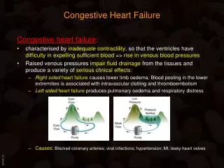
CSAS due to Cheyne Stokes Respiration • Cheyne-Stokes respiration (CSR) is characterized by an absence of air flow and respiratory effort followed by hyperventilation in a crescendo-decrescendo pattern. • CSR most often occurs in patients with congestive heart failure (CHF). • The prevalence is estimated to be approximately 30% to 40% in patients with CHF. • This respiratory pattern can also be seen in patients with stroke or renal failure. • There is mounting evidence that CSAS/CSR may be an indicator of higher morbidity and mortality in CHF patients. • Effective treatment of CSAS/CSR might improve the outcome of CHF patients with CSAS/CSR.
CSAS Due to Medical Condition Not Cheyne Stokes • CSAS can occur in individuals with cardiac, renal, and neurological disorders but without a CSR pattern. • This category is referred to CSAS Due to Medical Condition Not Cheyne Stokes.
Complex sleep apnea • CompSA is defined as a form of CSA identified by the persistence or emergence of central sleep apneas or hypopneas upon exposure to CPAP or BPAP without a backup rate when obstructive events have disappeared. • These patients have predominantly obstructive or mixed apneas during the diagnostic portion of the study occurring 5/hr or more. • With use of CPAP or BPAP without a backup rate, they show a pattern of apneas and hypopneas that meets the definition of CSA.
Clinical Classification of AHF • The patient with AHF will usually present in one of six clinical categories. Pulmonary oedema may or may not complicate the clinical presentation. 1- Worsening or decompensated chronic HF (peripheral oedema / congestion): there is usually a history of progressive worsening of known chronic HF on treatment, and evidence of systemic and pulmonary congestion. Low BP on admission is associated with a poor prognosis. 2- Pulmonary oedema: patients present with severe respiratory distress, tachypnoea, and orthopnoea with rales over the lung fields. SPo2 is usually <90% on room air prior to treatment with oxygen. 3- Hypertensive HF: signs and symptoms of HF accompanied by high BP and usually relatively preserved LV systolic function. There is evidence of increased sympathetic tone with tachycardia and vasoconstriction. The patients present frequently with signs of pulmonary congestion without signs of systemic congestion. The response to appropriate therapy is rapid, and hospital mortality is low.
Clinical Classification of AHF 4- Cardiogenic shock: is defined as evidence of tissue hypoperfusion induced by HF after adequate correction of preload and major arrhythmia. Typically, cardiogenic shock is characterized by reduced systolic blood pressure (SBP<90 mmHg or a drop of mean arterial pressure >30 mmHg) and absent or low urine output (< 0.5 mL/kg/h). Evidence of organ hypoperfusion and pulmonary congestion develop rapidly. 5- Isolated right HF: is characterized by a low output syndrome in the absence of pulmonary congestion with increased jugular venous pressure, with or without hepatomegaly, and low LV filling pressures . 6- Acute coronary syndrome (ACS) and HF: Approximately 15% of patients with an ACS have signs and symptoms of HF . Episodes of acute HF are frequently associated with or precipitated by an arrhythmia (bradycardia, Atrial fibrillation, Ventricular tachycardia).
Non-invasive ventilation In AHF • NIV with positive end-expiratory pressure (PEEP) should be considered as early as possible in every patient with acute cardiogenic pulmonary oedema and hypertensive AHF as it improves clinical parameters including respiratory distress. • NIV with PEEP improves LV function by reducing LV afterload. • NIV should be used with caution in cardiogenic shock and right ventricular failure.
Acute Cardiogenic Pulmonary Edema • Acute cardiogenic pulmonary edema is the first cause of acute respiratory distress worldwide. • The initial management of patients with ACPE address theABCs (airways, breathing, circulation). • Oxygen should be adminstered to all patiients to keep oxygen >90%. • Any associated arrhythmia or infarction should be treated appropriately .
Acute Cardiogenic Pulmonary Edema Medical treatment of ACPE: • Reduction of pulmonary venous return (preload reduction) • Reduction of systemic vascular resistance (afterload reduction), and, in some cases, • Inotropic support
Acute Cardiogenic Pulmonary Edema • Preload reduction decreases pulmonary capillary hydrostatic pressure and reduces fluid transudation into the pulmonary interstitium and alveoli. • Afterload reduction increases cardiac output and improves renal perfusion, which allows for diuresis in the patient with fluid overload. • Patients with severe LV dysfunction or acute valvular disorders present with hypotension. These patients may not tolerate medications to reduce their preload and afterload. Therefore, inotropic support is necessary in this subset of patients to maintain adequate blood pressure. • Patients who remain hypoxic despite supplemental oxygenation and patients who have severe respiratory distress require ventilatory support in addition to maximalmedical therapy.
Acute Cardiogenic Pulmonary Edema • In patients with acute respiratory failure, standard treatment, including diuretics, nitroglycerin, morphine, and oxygen, may not be sufficient to reduce respiratory distress. • In this setting, noninvasive ventilation supportshould be initiated rapidly, with the main goals to 1-Improve oxygenation, 2- Avoid invasive ventilation, and 3- Permit a sufficient period for medical therapy to decrease pulmonary vascular congestion.
Acute Cardiogenic Pulmonary Edema • Methods of oxygen delivery 1-Face mask 2- Noninvasive pressure-upport ventilation (which includes BiPAP and CPAP), and 3-Intubation and mechanical ventilation • Which method is used depends on the presence of hypoxemia and acidosis and on the patient's level of consciousness. For example, intubation and mechanical ventilation may become necessary in cases of persistent hypoxemia, acidosis, or altered mental status .
What should be the interface? oronasal masks - general advantage • Best suited for less cooperative patients • Better in patients with a higher severity of illness • Better for patients with mouth-breathing or pursed-lips breathing • Better in edentulous patients • Generally more effective ventilation oronasal masks- cautions, disadvantages • Claustrophobic • Hinder speaking and coughing • Risk of aspiration with emesis
What should be the interface? Nasal masks - general advantages • Best suited for more cooperative patients • Better in patients with a lower severity of illness • Not claustrophobic • Allows speaking, drinking, coughing, and secretion clearance • Less aspiration risk with emesis • Generally better tolerated Nasal masks -cautions, disadvantages • More leaks possible (eg, mouth-breathing or edentulous patients) • Effectiveness limited in patients with nasal deformities or blocked nasal passages
What should be the interface? • It cannot be said that any interface is clearly superior to another in terms of important outcomes such as intubation rate or mortality. • An oro-nasal interface may be more effective and better tolerated than the nasal interface for patients with acute respiratory failure. Thus, a sensible approach would be to start with an oronasal mask for most patients with acute respiratory failure, and switch to a nasal mask if prolonged use is contemplated. • Whichever mask is chosen, a comfortable fit is of paramount importance, and thus using a mask of proper size, not strapping the headgear too tightly, and using wound care tape on the bridge of the nose are important considerations to avoid pressure ulcers.
Does the type of ventilator make any difference? • NIV can be delivered through ventilators designed for invasive mechanical ventilation (‘‘critical care ventilators’’),and portable devices. • Critical care ventilators are less leak tolerant and are thus likely to sound alarms more inappropriately. But the monitoring capabilities and presence of oxygen blenders make it superior to portable devices. • On the other hand, the portable ventilators are more leak tolerant and less likely to sound alarms inappropriately than the critical care ventilators. However, they may promote rebreathing by virtue of their single inspiratory and expiratory tubing (minimized by assuring adequate expiratory pressure and expiratory ports over the nasal bridge)
Does the type of ventilator make any difference? • Most of the portable ventilators do not have an oxygen blender and supplemental oxygen is usually given by adding it into the mask or the circuit. • Continuous pulse oximetry is required to monitor oxygenation when using this device in patients with ACPE. • Comparisons of the two devices show that the portable device performs as well as the critical care ventilators. • Recently, ventilators that deliver either invasive ventilation or NIV have been designed. When in the non-invasive mode, they are more leak tolerant and use only the alarms essential for the operation of NIV. • The choice of CPAP versus BPAP can be dictated by local experience and patient preference, although BPAP may have some additional benefit to those with hypercapnic acidosis.
Where are these patients best treated? • Patients with severe ACPE requiring NIV need to be triaged to an environment with adequate nurse-patient ratio, and continuous electrocardiographic and pulse oximetry monitoring facilities.
Do all patients with ACPE require NIV? • Consider NIV early when treating patients with severe ACPE. • Several studies suggest that NIV is associated with decreased length of stay in the ICU, decreased need for mechanical ventilation, and decreased hospital costs. • A few clinical trials showed that early and prehospital NIV treatment by paramedics is safe and associated with faster improvement of oxygen saturation. • However, the mortality and the need for intensive care did not differ between the patients who were treated with NIV and those who were treated with a Venturi face mask in most of those studies.
AHF treatment strategy according to systolic blood pressure.
Venturi face mask (Air entrainment mask) The colour of the device reflects the delivered oxygen concentration: 24%: blue; 28%: white; 31%: orange; 35%: yellow; 40%: red; 60%: green.