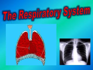
The Respiratory System
690 likes | 840 Vues
The Respiratory System. Intended Learning Outcomes. Pages 270-280. Identify and give the functions for each of the following: Larynx,Trachea , Bronchi, Bronchioles, Alveoli, Diaphragm & ribs, Pleural membranes, and Thoracic cavity.

The Respiratory System
E N D
Presentation Transcript
Intended Learning Outcomes Pages 270-280 • Identify and give the functions for each of the following: • Larynx,Trachea, Bronchi, Bronchioles, Alveoli, Diaphragm & ribs, Pleural membranes, and Thoracic cavity. • Compare and contrast the mechanics of the processes of inhalation and exhalation . • Explain the relationship between the structure and function of the alveoli. • Explain the roles of the cilia and mucous in the respiratory tract. • Describe the interaction of the lungs, pleural membranes, ribs, and diaphragm in the breathing process. • Explain the roles of carbon dioxide and hydrogen ions in stimulating the breathing centre in the medulla oblongata. • Describe the exchange of carbon dioxide and oxygen during internal and external respiration. • Distinguish between the transport of carbon dioxide and oxygen in the blood by explaining the roles of oxyhemoglobin, carboxyhemoglobin, reduced hemoglobin, and bicarbonate ions.
Vocabulary _____ Alveoli _____ Bicarbonate ions _____ Breathing _____ Bronchi _____ Bronchioles _____ Carbaminohemoglobin(HbCO2) _____ Carbon dioxide _____ Carbonic anhydrase _____ Carboxyhemoglobin (HbCO2) _____ Cartilage _____ Cellular respiration _____ Chemoreceptors _____ Cilia _____ Diaphragm _____ Epiglottis _____ Esophagus _____ Exhalation/Expiration _____ External respiration _____ Hemoglobin _____ Hydrogen ions _____ Inspiration/Inhalation _____ Intercostal muscles _____ Internal respiration _____ Larynx _____ Lipoproteins (surfactant) _____ Medulla oblongata _____ Mucous _____ Nasal sinus _____ Nose hairs _____ Oxyhemoglobin (HbO2) _____ Pharynx _____ Pleural membrane _____ Pneumothorax _____ Pulmonary capillaries _____ Reduced hemoglobin (HHb) _____ Sinus _____ Stretch receptors _____ Surface tension _____ Thoracic cavity _____ Trachea _____ Vocal cords
Interesting Facts The right lung is slightly larger than the left. Hairs in the nose help to clean the air we breathe, as well as warming it. The highest recorded "sneeze speed" is 165 km per hour. The surface area of the lungs is roughly the same size as a tennis court. Each red blood cell has about 200-250 million Hemoglobin molecules.
More Interesting Facts The capillaries in the lungs would extend 1,600 km if placed end to end. We lose half a litreof water a day through breathing. This is the water vapour we see when we breathe onto glass. A person at rest usually breathes between 12 and 15 times a minute. The breathing rate is faster in children and women than in men. Lung breathing probably evolved about 400 million years ago.
The Nasal Sinus • The Nasal Sinus is surrounded by a lot of capillary beds and mucous glands. Because it is one of the major entry ways into the body it has many things to help keep us safe • Nose hairs: with the aid of mucous, these hairs filter and trap debris. The debris that is trapped in this manner is discharged through the nose. • There are many white blood cells here to recognize and destroy foreign objects. • Histamines are released here as an allergic response when foreign irritants are encountered. This causes runny nose.
Pharynx/Throat This is the common passageway for air and food
Epiglottis This is a flap of tissue that covers the top of the trachea when swallowing to ensure that food enters the esophagus and not the lungs.
Larynx When the epiglottis is opened, the air is able to pass through the larynx (voice box) and into the trachea. The larynx contains the vocal cords (two tendons that adjust the pitch of sounds according to how taut they are).
When a guy goes through puberty, his vocal chords and voice box (larynx) grow larger, and begins to stick out at the front of the throat. This lump is called the Adam's Apple. Male vocal cords: 17 mm & 25 mm in length. Female vocal cords: 12.5 mm & 17.5 mm in length.[1
Trachea This is the windpipe. This passageway is held open by the presence of C-shaped rings of cartilage. This is a protective adaptation. The trachea conducts air into the bronchi.
Cilia and mucus filter the air as it moves through the trachea. • The mucous traps the dirt and other particles, and the cilia push it to the back of the throat so we swallow it into our digestive system Cilia with pollen trapped by mucous.
Conditioning of the Air • Several things happen to the air on its way to the alveoli. It is: • Adjusted to Body Temperature: By the time it arrives at the alveoli the air has been in contact with many tissues and is 37o C. • Adjusted to 100% humidity. As inhaled air passes over the mucous passageways, it becomes saturated with water. • Cleansed of debris in a 2 part process. • Nose hairs and mucous in the nasal passageways. • Mucous and Cilia in Trachea and bronchi. *Note: cilia do not filter!
Bronchi The trachea splits into two bronchi and takes the air into each lung. These branches also have cartilage around them, for the same reason. The bronchi conducts air into smaller branching passageways called bronchioles.
Bronchioles • The bronchioles are branching passageways that carry air to its ultimate destination, the alveoli.
Alveoli These are the blind sac-like endings at the end of the bronchioles. There are approx. 700,000 alveoli in the human lung. This is the site of gas exchange. O2 leaves the alveoli and moves into the blood to be taken around the body. CO2 does the opposite and is breathed out.
Why are they so special? • NUMEROUS: Each adult lung contains millions of alveoli. This provides lots of surface area for the gases to be exchanged. • THIN WALLS: The walls of alveoli are only one cell thick. • STRETCH RECEPTORS: They have stretch receptors that signal when the alveoli are full enough (stretched). They send a message to the brain to start exhalation.
Why are they so special? • MOIST: They are very moist and this helps gas exchange. • VERY RICH BLOOD SUPPLY: They have a close association with many blood capillaries so oxygen and carbon dioxide can be exchanged efficiently. • LINED WITH A LAYER OF LIPOPROTEINS (surfactant) on their inner surface. This helps to maintain surface tension, thus preventing them from collapsing and sticking together during exhalation.
Diaphragm This is a sheet of muscle that separates the chest cavity from the abdominal cavity. When you inhale it moves down. When you exhale it moves up.
Ribs and Muscles • These are the bones that are connected to the vertebral column and sternum. • These are INTERCOSTAL muscles between the ribs, which help to move the ribs… • Up and out when we inhale. • Down and in when we exhale.
MEDULLA OBLONGATA Our control of the breathing process is only voluntary to a point. The medulla oblongata of the brain is sensitive to the concentration of carbon dioxide and hydrogen ions in the blood. When the concentrations of H+ and CO2reach a critical level, the breathing center in the medulla oblongata is stimulated and sends nerve impulses to the diaphragm and the intercostal muscles.
TO INHALE When the brain realizes there is too much CO2in our blood, it sends a message to the rib muscles and diaphragm to contract. The ribs move up and out, the diaphragm moves down. This creates more space in the lungs (negative air pressure) and air rushes in to fill that space. This is called inhalation.
TO EXHALE When the alveoli get too stretched (full of air), they send a message to the brain to stop inhaling. The brain tells the muscles to relax and the ribs move back down and in, while the diaphragm moves back up. This decreases the amount of space in the lungs and the air is pushed out. This is called exhalation.
See a Working Respiratory System:http://www.smm.org/heart/lungs/breathing.htm
Pleural Membranes • These are membranes that enclose the lungs. • The outer pleural membrane sticks closely to the walls of the chest & the diaphragm. • The inner pleural membrane is stuck to the lungs. The two lie very close to each other. The pleura allows the surface of the lungs to slide over the body wall easily, without abrasion.
Pleural Membranes The pleura seals off the thoracic cavity so when the lungs inflate, a negative air pressure is created and this causes air to rush in. These membranes also stick the lungs to the chest cavity walls, so when the ribs move out, so do the lungs.
Pneumothorax A puncture to the chest wall, piercing the pleural membrane (even without damaging the lung itself), will result in a pneumothorax, or collapse of the lung. In a situation like this, the negative pressure effectively draws air in through the puncture wound, putting pressure on the surface of the lung instead of inside it and the lung collapses.
Respiratory System • Respiration is the set of processes involved with the conduction of oxygen to the tissues and the removal of the waste product CO2. • There are four aspects to respiration: • 1. Breathing: the inspiration and expiration of air. • 2. External Respiration: gas exchange at the alveoli. • Internal Respiration: gas exchange at the tissues. • Cellular Respiration: mitochondria turn O2 and glucose into CO2 and H2O and ATP energy.
External Respiration • Happens at the lungs. • It is the diffusion of O2 into the pulmonary capillaries (blood) and the diffusion of CO2 and water into the alveoli to be exhaled with the air.
External Respiration Because there is a lot of CO2 returning to the lungs, and not very much in the alveoli, the CO2 diffuses from [H] to [L] down its concentration gradient and moves into the alveoli to be breathed out. Because there is a lot of O2 in the fresh air in the alveoli, and not much in the deO2 blood, the oxygen diffuses from [H] to [L] down its concentration gradient and into the blood.
External Respiration Conditions in the blood at the alveoli are: Under these conditions, hemoglobin lets go of CO2and starts to love oxygen. As it leaves the lungs, 99% of hemoglobin is occupied with oxygen it is called oxyhemoglobin. Hemoglobin transports oxygen to the tissue cells. Basic: pH of ~7.4 Cool: ~37o C Low (negative) pressure O2 + Hb HbO2
Internal Respiration • Happens at the tissues. • It is the diffusion of O2 into the tissue cells, and the diffusion of CO2 and water into the blood capillaries. • The CO2 is then returned to the heart and sent to the lungs to be removed during exhalation.
Internal Respiration CO2 O2
Internal Respiration Conditions in the blood at the tissues are: Under these conditions, hemoglobin lets go of O2and starts to love CO2 and H+. The oxygen diffuses into the tissue spaces along with the water that is forced from the plasma due to blood pressure. Acidic: pH of ~7.3 Warm: ~38o C High pressure HbO2 Hb + O2
Cellular Respiration CO2 + H2O + O2 + GLUCOSE
Internal Respiration (con’t) At the venule end of the capillary bed, when water is drawn back into the blood by osmotic pressure, CO2 enters the blood. Carbon dioxide can be transported in three ways: 1. Dissolved gas, 2. Carboxyhemoglobin (HbCO2), 3. Bicarbonate ion (HCO3-)
Internal Respiration (con’t) Because the hemoglobin now loves the CO2, they join to form carboxyhemoglobin. CO2 also joins with water to make the bicarbonate ion. There is an enzyme in the red blood cells called CARBONIC ANHYDRASE which catalyzes this reaction. CO2 + Hb HbCO2 CO2 + H2O H2CO3 HCO3- + H+ Carbonic anhydrase Carbonic anhydrase
Internal Respiration (con’t) CO2 + H2O H2CO3 HCO3- + H+ Carbonic anhydrase Carbonic anhydrase The extra hydrogen ion from the water is now free. This is BAD as it is ACIDIC and can eat through the blood vessel walls. So, hemoglobin acts as a buffer and joins with the hydrogen ion to make reduced hemoglobin. H+ + Hb HHb


