-Hemolytic Streptococci
-Hemolytic Streptococci. Ali Somily MD,FRCPC,D(ABMM). G+ve cocci in chains and/or pairs Colonize MM(Resp.,GIT& GUT) Catalase –ve Ferment CHO lactic acid. Grouped by either A.phenotypic Hemolysis( ,ß or ) Lancefield antigen Cell wall CHO A,B,C,D,Fand G ect Or B.Genotypic.

-Hemolytic Streptococci
E N D
Presentation Transcript
-Hemolytic Streptococci Ali Somily MD,FRCPC,D(ABMM)
G+ve cocci in chains and/or pairs Colonize MM(Resp.,GIT& GUT) Catalase –ve Ferment CHOlactic acid Grouped by either A.phenotypic Hemolysis(,ß or ) Lancefield antigen Cell wall CHO A,B,C,D,Fand G ect Or B.Genotypic Introduction
-Hemolytic Streptococci • Partial hemolysis of blood • Green zoon around the colony • Examples: • S.Pneumoniae • S.Viridans • S.Bovis
VIRULANCE VACTORS Capsule Autolysin Pneumolysin CLINICAL PRESENTATION NF Resp.20-40%/pharynx Inhalation/droplets STREPTOCOCCUS PNEUMONIAE
Primary infection CAPalveoli Blood Endocarditis Meningitis Localized Sinusitis O.M Secondary Infection Non-capsulated Opportunistic infection Lungs only Impair or poor ciliary activity Viral, Smoking, dust Risk factor Hyposplenism Splenectomy Asplenia Sickle Cell Diseases Liver disease Hypogammaglobinaemia Alcoholism Cigarette smoking Malnutrition CLINICAL PRESENTATION
Blood,CSF,Sputum& swab/aspirate DIRECT SMEAR G+ve diplococci Lancet shape Capsulated halo (antiphagocytic)/pathogenic Diagnosis
Quellung test(AB’s swelling of capsule) Diagnosis
CULTURE BAP; 5-10%CO2 -hemolytic Mucoid (capsule)SR Concave (punched out/collapse) IDENTIFICATION Bile solubility (NaDC) Optochin S (disk 5g&6mmzoon>=14 mm) 80 serotype(vaccine) capsular structure STREPTOCOCCUS PNEUMONIAE
Treatment &Prevention • TTT • Penicillin(↑R recently)PBP • Ceftriaxon +/-Vancomycin or Rifampicin • Vaccination • Polsaccharide capsule • Conjugate vaccine • Indication • Children • SCD • Splenectomised patient • HIV • Elderly • Cardiopulmonary and renal diseases
Mitis Mutans Salvarius Angionosis Grouping of viridans streptococci
CLINCAL PRESENTATION NF (GIT,UGT&Oral cavity) Opportunistic organisms Dental caries Endocarditis VIRIDANS STREPTOCOCCI
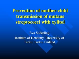
Formation of dental plaque by Streptococcus mutans bacteria adhere to the tooth by a protein on the cell surface, grow and synthesize a dextran capsule binds the bacteria to the enamel and forms a biofilm 300-500 cells of thickness bacteria can cleave sucrose to glucose + fructose glucose is polymerized into an extracellular dextran polymer that cements the bacteria to tooth enamel and becomes the matrix of plaque this dextran slime can be depolymerized to glucose for use as a carbon source, resulting in the production of lactic acid within the plaque that decalcifies the enamel and leads to dental caries Example of a biofilm
Damage ,prosthetic valve Predisposing factor RHD CHD Mitral valve prolapse Degenerative H diseases PV Pathogenesis Dextran adhere to the damaged valve vegetation(fibrin,bacteria & platelets) Symptoms Fever Murmurs Immunological manifestations O,P,S SBE 2-5 Wks Diagnosis Blood culture ECHO Endocarditis
DIRECT SMEAR G+ve cocci in chains CULTURE Can be OR rarely ß hemolytic Most lack distinct LA IDENTIFICATION Urea VP( acetoin production) Arginine hydrolysis Esculin hydrolysis CHO ferementation Microbiology
Introduction • Fecal strep separated genus/by molecular • Harsh conditionAbiquitous/soil,water,plants, GIT, GU human • 15 Spp/E.faecalis80-90% of clinical isolate • Colonization/infection
NC BACTEREMIA 1/3 NC UTI 16% WOUND ENDOCARDITIS CNS PNEUMONIA (rare) ELDERLY I’C TTT Vancomycin VRE (Van A,B&C ect) Plasmid/chromosomal High/low level VDE Direct smear Short chain/pairs/coccobaccilary CLINICAL PRESENTATION
CULTURE Facultative anaerobs OR hemolysis Catalase weak +ve Group D IDENTIFICATION Growth B/W 10-42 Co 6.5% NaCl Esculin hydrolysis(40%)bile salt Pyrrolidonyl arylamidase(PYR)+ve Leucine arylamidase(LAP)+ve Motility ,Pigmentation,arabinose and methyl --D-glucopyranoside
All Group D Enterococcus S.Bovis Bile esculin
BOVIS GROUPNew NameStreptococcus gallolyticus(Group D Streptococci)
CLINICAL Human intestine and animals Endocarditic Septicemia Link to colon cancer Stool isolation Two biotypes I&II Mannitol and glucan I DIRECT SMEAR G+ve cocci in chains BOVIS GROUP
CULTURE OR on BAP IDENTIFICATION LG group D Differed from viridans Growth in 40% bile salt Hydrolyze esculin
S.bovis Unable to grow in 45 Co 6.5% salt @ 37dC PYR –ve S to penicillin and cephalosporins R to vancomycin(rare) Enterococcus Able to grow in 45 Co 6.5% salt @ 37Co PYR +ve S to ampicillin How you differentiate S. bovis from enterococcus?