Bone tissue
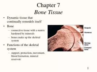
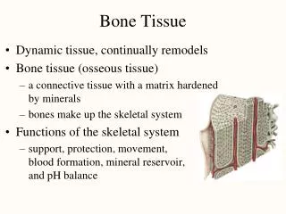
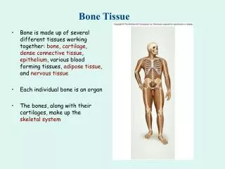
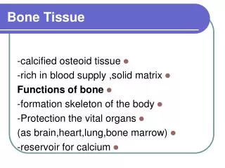
Bone tissue. Macroscopic features. Diaphysis – bone shaft – sight of compact bone Epiphyses – sight of spongy bone Bone marrow Red is sight of hematopoiesis (red blood cell formation) Yellow is fat stores Periosteum – outer surface Endosteum – lines inner surfaces Compact bone

Bone tissue
E N D
Presentation Transcript
Macroscopic features • Diaphysis – bone shaft – sight of compact bone • Epiphyses – sight of spongy bone • Bone marrow • Red is sight of hematopoiesis (red blood cell formation) • Yellow is fat stores • Periosteum – outer surface • Endosteum – lines inner surfaces • Compact bone • Spongy bone
Microscopic features • Lacunae • Small pockets containing osteocytes • Lamellae • Narrow layers of calcified matrix • Canaliculi • Small channels that radiate through the matrix, interconnecting lacunae and connecting them to nearby blood vessels.
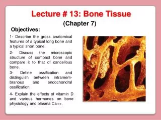
Compact bone (microscopic) • Osteon or Haversian system – basic unit • Osteocytes are arranged in circular layers around a Haversian canal that contains blood vessels and nerves. • Layer of compact bone covers bone surfaces everywhere except inside joint capsules • Compact bone usually found where stresses come from a limited range of directions.
Spongy bone (microscopic) • No osteons and different lamellar arrangement than compact bone. • Lamellae form plates called trabeculae. • Spongy bone is found where bones are not heavily stressed or where stresses arrive from many directions. • Spongy bone is much lighter than compact.
Cells in bone Osteocytes - mature bone cells - maintain normal bone structure by recycling calcium salts and by assisting in repairs
Cells in bone Osteoclasts - giant cells with 50 or more nuclei - acids and enzymes secreted by osteoclasts dissolve bony matrix and release stored minerals.
Cells in bone Osteoblasts - produce new bone matrix and promote deposition of calcium salts in matrix. - immature, matrix-depositing bone cells, responsible for production of new bone (osteogenesis).
Bone formation and growth • Skeletal growth begins about 6 weeks after fertilization and continues until about age 25. • Ossification – during development, cartilage or other connective tissues are replaced by bone.
Endochondral ossification • Most skeletal bone forms through this process • Ossification of existing hyaline cartilage • Cartilage between shaft and epiphysis does not completely fill with bone because the epiphyseal plates (growth plate) on the ends continue to enlarge, increasing the length of developing bone.
Requirements for normal bone growth • Must have a reliable source of minerals, especially calcium salts. • Diet must provide adequate amounts of calcium and phosphate and the body must be able to absorb and transport these minerals to sites of bone formation. • When blood calcium levels drop, parathyroid hormone is released causing calcium to be released from bone.
Vitamin D • plays a role in normal calcium metabolism • Can be obtained from dietary supplements or manufactured by epidermal cells exposed to UV radiation.
Vitamin A and C • Essential for normal bone growth and maintenance. • Vitamin C deficiency can lead to Scurvy with weak brittle bones.