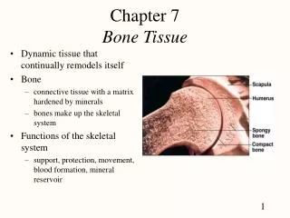
Bone Tissue
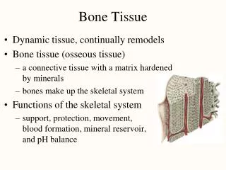
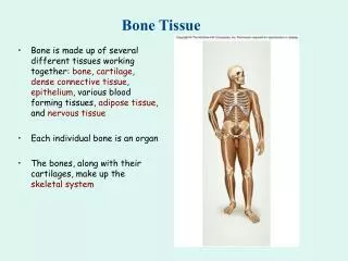
Bone Tissue. Bone is made up of several different tissues working together: bone , cartilage , dense connective tissue , epithelium , various blood forming tissues, adipose tissue , and nervous tissue Each individual bone is an organ

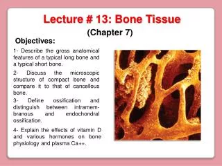
Bone Tissue
E N D
Presentation Transcript
Bone Tissue • Bone is made up of several different tissues working together: bone, cartilage, dense connective tissue, epithelium, various blood forming tissues, adipose tissue, and nervous tissue • Each individual bone is an organ • The bones, along with their cartilages, make up the skeletal system
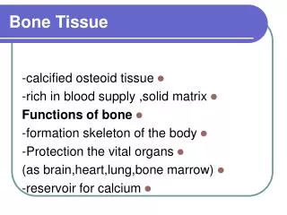
Functions of Bone • Supporting & protecting soft tissues • Attachment site for muscles making movement possible • Storage of minerals, especially calcium & phosphate (mineral homeostasis) • Blood cell production occurs in red bone marrow (hemopoiesis & EPO) • Energy storage in yellow bone marrow • Gives pirates something to put on their flags
Anatomy of a Long Bone • Diaphysis = shaft (compact bone) • Epiphysis = one end of a long bone (spongy bone) • Metaphyses are the areas between the epiphysis and diaphysis and include the epiphyseal plate in growing bones • Articular cartilage over joint surfaces acts as friction reducer & shock absorber • Medullary cavity = marrow cavity
Anatomy of a Long Bone • Endosteum = lining of marrow cavity • Periosteum = tough membrane covering bone but not the cartilage • Fibrous layer = dense irregular connective tissue • Osteogenic layer = bone cells & blood vessels that nourish or help with repairs
Anatomy of a Flat Bone • Internal and external surfaces formed of compact bone • Middle layer is spongy bone and red bone marrow • Fractures may break the compact bone layer but leave the spongy bone relatively unharmed
General Histology of Bone • Bone is a type of connective tissue as seen by widely spaced cells separated by matrix • Matrix of 25% water, 25% collagen fibers & 50% crystalized mineral salts • ~85% hydroxyapatite, a crystalized calcium phosphate salt • Mineral salts are deposited in a framework of collagen fibers, a process known as mineralization or calcification • Hardness of bone depends on mineral salts, flexibility depends on collagen fibers (“like fiberglass”)
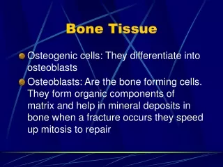
Bone Cells • OsteogenicCells: Undifferentiated cells • can divide to replace themselves & can become osteoblasts • found in inner layer of periosteum and endosteum • Osteoblasts: Form matrix & collagen fibers but can’t divide • Osteocytes: Mature cells that no longer secrete matrix, now trapped in the matrix they secreted • Osteoclasts: Huge cells from ~50 fused monocytes (WBC) • function in bone resorption at surfaces such as endosteum
Compact or Dense Bone • Looks like solid hard layer of bone • Makes up the shaft of long bones and the external layer of all bones • Resists stresses produced by weight and movement
Compact or Dense Bone • Compact bone is arranged in units called osteons or Haversian systems • Osteons contain blood vessels, lymphatic vessels, nerves, and osteocytes along with the calcified matrix • Osteons are aligned in the same direction along lines of stress, which can slowly change as the stresses on the bone change
Histology of Compact Bone • Osteon is concentric rings (lamellae) of calcified matrix surrounding a vertically oriented blood vessel • Osteocytes are found in spaces called lacunae • Osteocytes communicate through canaliculi filled with extracellular fluid that connect one cell to the next cell • Interstitial lamellae represent older osteons that have been partially removed during tissue remodeling
Structure and Histology of Spongy Bone • Generally does NOT contain osteons • Latticework of thin plates of bone called trabeculae oriented along lines of stress • Provides strength with little weight • Spaces in between struts of trabeculae are filled with red marrow where red blood cells develop • Central canals not needed because no osteocyte is far from the marrow • Found in epiphyses of long bones and inside flat bones such as the hipbones, sternum, sides of skull, and ribs No Osteons
Blood and Nerve Supply of Bone • Periosteal arteries • Supply periosteum • Nutrient arteries • Enter through nutrient foramina • Supply compact bone of diaphysis & red marrow • Metaphyseal & epiphysealarteries • Supply red marrow & bone tissue of epiphyses
Bone Formation • All embryonic connective tissue begins as mesenchyme • Bone formation is termed osteogenesis or ossification and begins when mesenchymal cells provide the template for subsequent ossification • Two types of ossification occur • Intramembranous ossification is the formation of bone directly from or within fibrous connective tissue membranes • Flat bones of the skull • Thin layers of compact bone with spongy bone in the middle • Endochondrial ossification is the formation of bone from hyaline cartilage models, forming most of the bones in the body • Two growth centers: primary & secondary • Two types of growth: interstitial & appositional
Intramembranous Bone Formation • Mesenchyme condenses into sheets • Osteoblasts secrete matrix & become osteocytes, periosteum forms • Trabeculae form, creating spongy bone • Calcification converts surfaces to compact bone
Endochondral Bone Formation • Cartilage model forms • Formation of periosteum and theprimary ossification center in the diaphysis • Vascular invasion, formation of medullary cavity • Formation of secondary ossification center in epiphysis • Epiphyseal plate forms, bone grows in length from this plate • Epiphyseal plate closes to form epiphyseal line, bone stops growing
Bone Growth in Length • Epiphyseal plate or cartilage growth plate • Cartilage cells are produced by mitosis on epiphyseal side of plate • Cartilage cells are destroyed and replaced by bone on diaphyseal side of plate • Between ages 18 to 25, epiphyseal plates close • Cartilage cells stop dividing and bone replaces the cartilage, forming the epiphyseal line • Growth in length stops at age ~25
Zones of Growth in Epiphyseal Plate • Zone of reserve cartilage • Anchors growth plate to bone • Zone of cell proliferation • Rapid cell division (stacked coins) • Zone of cell hypertrophy • Mitosis stops, cells enlarge • Zone of calcification • Cartilage matrix calcifies • Zone of bone deposition • Osteoclasts break down lacunae • Osteoblasts & capillaries move in to create bone over calcified cartilage
Bone Growth in Thickness • Only by appositional growth at the bone’s surface • Periosteal cells differentiate into osteoblasts and form bony ridges and then a tunnel around periosteal blood vessel • Concentric lamellae fill in the tunnel to form an osteon
Factors Affecting Bone Growth • Nutrition • Adequate levels of minerals and vitamins • Calcium and phosphorus for bone growth • Vitamin C for collagen formation • Vitamin K and Vitamin B12 for protein synthesis • Sufficient levels of specific hormones • During childhood need insulinlike growth factor (IGF) • Promotes cell division at epiphyseal plate • Human growth hormone (hGH), thyroid hormones, & insulin • Sex steroids at puberty • Estrogen and testosterone, stimulate sudden growth and modifications of the skeleton to create the male and female forms Testosterone
Hormonal Abnormalities in Bone Growth • Oversecretion of hGH during childhood produces giantism • Oversecretion of hGH during adulthood results in acromegaly • From the Greek akros = high and megalos = large • Individuals with acromegaly have unusually large facial features, large hands, large feet, and wide tooth spacing • Usually caused by a benign tumor of the pituitary gland • Today acromegaly can be treated with drugs or surgery • Undersecretion of hGH or thyroid hormone during childhood produces short stature • Both men or women that lack estrogen receptors on cells grow taller than normal • estrogen is responsible for closure of growth plate
Hormonal Abnormalities in Bone Growth • Worked as a journalist • Originally given a bit part in a movie because of his looks • Inducted into his HS football team’s Hall of Fame • Rondo Hatton (1894 - 1946) • Suffered from acromegaly • “Star” of “fright films” of the 1930’s & 1940’s • Immortalized by the Rondo Hatton Classic Horror Awards • The Brute Man • Jungle Captive • House of Horrors • The Creeper
Hormonal Abnormalities in Bone Growth • Richard Kiel (1939 -) • “Jaws” in James Bond films • Also in Happy Gilmore & other films
Mineral Homeostasis • Bone is a storehouse for calcium and phosphate • Phosphate is important in many physiological processes • Needed for ATP • Component of DNA, RNA • Forms phospholipid bilayer • The body’s pH buffering system • Calcium is one of the most important regulatory elements in the body • Needed for muscle contraction • Needed for release of neurotransmitter from synaptic end bulbs • Excess blood calcium is hypercalcemia (muscle weakness) • Insufficient blood calcium is hypocalcemia (muscle spasms)
Mineral Homeostasis • Calcitriol is a form of Vitamin D produced by the skin, liver, & kidney • Raises blood calcium by promoting absorption in small intestine & from bones • Calcitonin is secreted by the thyroid gland • Lowers blood calcium by stimulating osteoblasts & inhibiting osteoclasts • Parathyroid hormone (PTH) is secreted by the parathyroid glands • Raises blood calcium by stimulating osteoclasts & promoting resorption
Mineral Homeostasis • Regulation of blood calcium provides a good demonstration of negative feedback loops • Hypercalcemia is excess Ca2+ in the blood • Hypocalcemia is insufficient Ca2+ in the blood
Repair of a Fracture • Formation of fracture hematoma • Damaged blood vessels produce clot in 6-8 hours, bone cells die • Inflammation brings in phagocytic cells for clean-up duty • New capillaries grow into damaged area • Formation of fibrocartilagenous callus • Fibroblasts invade the fracture site & lay down collagen fibers • Chondroblasts produce fibrocartilage to span the broken ends of the bone
Repair of a Fracture • Formation of bony callus • Osteoblasts secrete spongy bone that joins 2 broken ends of bone • Lasts 3-4 months • Bone remodeling • Compact bone replaces the spongy bone in the bony callus • Surface is remodeled back to normal shape
Exercise And Bone Tissue • Within limits, bone has the ability to alter its strength in response to mechanical stress by increasing deposition of mineral salts and production of collagen fibers (Wolff’s Law) • Removal of mechanical stress leads to weakening of bone through demineralization (loss of bone minerals) and collagen reduction • Reduced activity while in a cast • Astronauts in weightless environment • Bedridden person • Weight-bearing activities, such as walking or moderate weightlifting, help build and retain bone mass
Osteoporosis • Decreased bone mass resulting in porous bones • Those at risk • White, thin, menopausal females • Athletes who are not menstruating due to decreased body fat & decreased estrogen levels • People allergic to milk or with eating disorders whose intake of calcium is too low • Smoking, drinking, & family history increase risk • Prevention or decrease in severity • Adequate diet, weight-bearing exercise, & estrogen replacement therapy (for menopausal women) • Behavior when young may be most important factor