Bone tissue
Learn about bone tissue, its components, functions, and formation process. Explore the histology of bone tissue, cells involved, and types of bone structure. Study compact and spongy bone, their differences, and bone formation processes.

Bone tissue
E N D
Presentation Transcript
Bone tissue Online notes
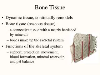
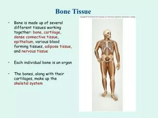
INTRODUCTION • Bone is made up of several different tissues working together: bone, cartilage, dense connective tissue, epithelium, various blood forming tissues, adipose tissue, and nervous tissue. • Each individual bone is an organ; the bones, along with their cartilages, make up the skeletal system.
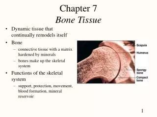
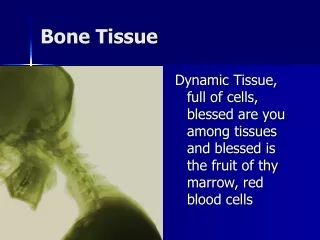
Chapter 6The Skeletal System:Bone Tissue • Dynamic and ever-changing throughout life • Skeleton composed of many different tissues • cartilage, bone tissue, epithelium, nerve, blood forming tissue, adipose, and dense connective tissue
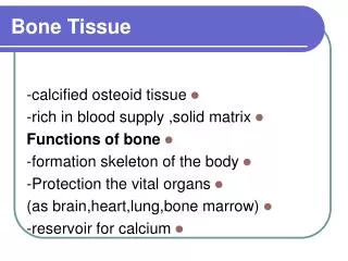
Functions of Bone • Supporting & protecting soft tissues • Attachment site for muscles making movement possible • Storage of the minerals, calcium & phosphate -- mineral homeostasis • Blood cell production occurs in red bone marrow (hemopoiesis) • Energy storage in yellow bone marrow
Anatomy of a Long Bone • diaphysis = shaft • epiphysis = one end of a long bone • metaphyses are the areas between the epiphysis and diaphysis and include the epiphyseal plate in growing bones. • Articular cartilage over joint surfaces acts as friction reducer & shock absorber • Medullary cavity = marrow cavity
Anatomy of a Long Bone • Endosteum = lining of marrow cavity • Periosteum = tough membrane covering bone but not the cartilage • fibrous layer = dense irregular CT • osteogenic layer = bone cells & blood vessels that nourish or help with repairs
bone cells.(Figure 6.2) • Osteogenic cells undergo cell division and develop into osteoblasts. • Osteoblasts are bone-building cells. • Osteocytes are mature bone cells and the principal cells of bone tissue. • Osteoclasts are derived from monocytes and serve to break down bone tissue.
HISTOLOGY OF BONE TISSUE • Bone (osseous) tissue consists of widely separated cells surrounded by large amounts of matrix. • The matrix of bone contains inorganic salts, primarily hydroxyapatite and some calcium carbonate, and collagen fibers. • These and a few other salts are deposited in a framework of collagen fibers, a process called calcification or mineralization. • The process of calcification occurs only in the presence of collagen fibers. • Mineral salts confer hardness on bone while collagen fibers give bone its great tensile strength.
Cells of Bone • Osteoprogenitor cells ---- undifferentiated cells • can divide to replace themselves & can become osteoblasts • found in inner layer of periosteum and endosteum • Osteoblasts--form matrix & collagen fibers but can’t divide • Osteocytes ---mature cells that no longer secrete matrix • Osteoclasts---- huge cells from fused monocytes (WBC) • function in bone resorption at surfaces such as endosteum
Matrix of Bone • Inorganic mineral salts provide bone’s hardness • hydroxyapatite (calcium phosphate) & calcium carbonate • Organic collagen fibers provide bone’s flexibility • their tensile strength resists being stretched or torn • remove minerals with acid & rubbery structure results • Bone is not completely solid since it has small spaces for vessels and red bone marrow • spongy bone has many such spaces • compact bone has very few such spaces
Compact Bone • Compact bone is arranged in units called osteons or Haversian systems (Figure 6.3a). • Osteons contain blood vessels, lymphatic vessels, nerves, and osteocytes along with the calcified matrix. • Osteons are aligned in the same direction along lines of stress. These lines can slowly change as the stresses on the bone changes.
Histology of Compact Bone • Osteon is concentric rings (lamellae) of calcified matrix surrounding a vertically oriented blood vessel • Osteocytes are found in spaces called lacunae • Osteocytes communicate through canaliculi filled with extracellular fluid that connect one cell to the next cell • Interstitial lamellae represent older osteons that have been partially removed during tissue remodeling
Spongy Bone • Spongy (cancellous) bone does not contain osteons. It consists of trabeculae surrounding many red marrow filled spaces (Figure 6.3b). • It forms most of the structure of short, flat, and irregular bones, and the epiphyses of long bones. • Spongy bone tissue is light and supports and protects the red bone marrow.
The Trabeculae of Spongy Bone • Latticework of thin plates of bone called trabeculae oriented along lines of stress • Spaces in between these struts are filled with red marrow where blood cells develop • Found in ends of long bones and inside flat bones such as the hipbones, sternum, sides of skull, and ribs. No true Osteons.
BONE FORMATION • All embryonic connective tissue begins as mesenchyme. • Bone formation is termed osteogenesis or ossification and begins when mesenchymal cells provide the template for subsequent ossification. • Two types of ossification occur. • Intramembranous ossification is the formation of bone directly from or within fibrous connective tissue membranes. • Endochondrial ossification is the formation of bone from hyaline cartilage models.
Growth in Length • To understand how a bone grows in length, one needs to know details of the epiphyseal or growth plate (Figure 6.7). • The epiphyseal plate consists of four zones: (Figure 6.7b) • zone of resting cartilage, • zone of proliferation cartilage, • zone of hypertrophic cartilage, and • zone of calcified cartilage The activity of the epiphyseal plate is the only means by which the diaphysis can increase in length. • When the epiphyseal plate closes, is replaced by bone, the epiphyseal line appears and indicates the bone has completed its growth in length.
Bone Growth in Length • Epiphyseal plate or cartilage growth plate • cartilage cells are produced by mitosis on epiphyseal side of plate • cartilage cells are destroyed and replaced by bone on diaphyseal side of plate • Between ages 18 to 25, epiphyseal plates close. • cartilage cells stop dividing and bone replaces the cartilage (epiphyseal line) • Growth in length stops at age 25
Zones of Growth in Epiphyseal Plate • Zone of resting cartilage • anchors growth plate to bone • Zone of proliferating cartilage • rapid cell division (stacked coins) • Zone of hypertrophic cartilage • cells enlarged & remain in columns • Zone of calcified cartilage • thin zone, cells mostly dead since matrix calcified • osteoclasts removing matrix • osteoblasts & capillaries move in to create bone over calcified cartilage
Growth in Thickness • Bone can grow in thickness or diameter only by appositional growth (Figure 6.8). • The steps in thes process are: • Periosteal cells differentiate into osteoblasts which secrete collagen fibers and organic molecules to form the matrix. • Ridges fuse and the periosteum becomes the endosteum. • New concentric lamellae are formed. • Osetoblasts under the peritsteum form new circumferential lamellae.
Factors Affecting Bone Growth • Nutrition • adequate levels of minerals and vitamins • calcium and phosphorus for bone growth • vitamin C for collagen formation • vitamins K and B12 for protein synthesis • Sufficient levels of specific hormones • during childhood need insulinlike growth factor • promotes cell division at epiphyseal plate • need hGH (growth), thyroid (T3 &T4) and insulin • sex steroids at puberty • At puberty the sex hormones, estrogen and testosterone, stimulate sudden growth and modifications of the skeleton to create the male and female forms.
Hormonal Abnormalities • Oversecretion of hGH during childhood produces giantism • Undersecretion of hGH or thyroid hormone during childhood produces short stature • Both men or women that lack estrogen receptors on cells grow taller than normal • estrogen is responsible for closure of growth plate
Bone Remodeling • Remodeling is the ongoing replacement of old bone tissue by new bone tissue. • Old bone is constantly destroyed by osteoclasts, whereas new bone is constructed by osteoblasts. • In orthodontics teeth are moved by brraces. This places stress on bone in the sockets causing osteoclasts and osteablasts to remodel the sockets so that the teeth can be properly aligned (Figure 6.2) • Several hormones and calcitrol control bone growth and bone remodeling (Figure 6.11)
Fracture and Repair of Bone A fracture is any break in a bone. • Fracture repair (Figure 6.10)involves formation of a clot called a fracture hematoma, organization of the fracture hematoma into granulation tissue called a procallus (subsequently transformed into a fibrocartilaginous [soft] callus), conversion of the fibrocartilaginous callus into the spongy bone of a bony (hard) callus, and, finally, remodeling of the callus to nearly original form.
Calcium Homeostasis & Bone Tissue • Skeleton is a reservoir of Calcium & Phosphate • Calcium ions involved with many body systems • nerve & muscle cell function • blood clotting • enzyme function in many biochemical reactions • Small changes in blood levels of Ca+2 can be deadly (plasma level maintained 9-11mg/100mL) • cardiac arrest if too high • respiratory arrest if too low
Hormonal Influences • Parathyroid hormone (PTH) is secreted if Ca+2 levels falls • PTH gene is turned on & more PTH is secreted from gland • osteoclast activity increased, kidney retains Ca+2 and produces calcitriol • Calcitonin hormone is secreted from parafollicular cells in thyroid if Ca+2 blood levels get too high • inhibits osteoclast activity • increases bone formation by osteoblasts