Statistical Parametric Mapping and Random Field Theory in Neuroimaging
Learn about Statistical Parametric Mapping (SPM) and Random Field Theory for accurate statistical inference in neuroimaging studies. Explore voxel-wise GLMs, Bayesian modeling, FWE correction, and spatial priors.

Statistical Parametric Mapping and Random Field Theory in Neuroimaging
E N D
Presentation Transcript
Statistical Parametric Mapping Will Penny Wellcome Trust Centre for Neuroimaging, University College London, UK LSTHM, UCL, Jan 14, 2009
Statistical Parametric Map Statistical Parametric Mapping Design matrix Image time-series Kernel Realignment Smoothing General linear model Random field theory Statistical inference Normalisation p <0.05 Template Parameter estimates
Outline • Voxel-wise General Linear Models • Random Field Theory • Bayesian modelling
Voxel-wise GLMs model specification parameter estimation Time hypothesis statistic Time Intensity single voxel time series SPM
Temporal convolution model for the BOLD response Convolve stimulus function with a canonical hemodynamic response function (HRF): HRF
General Linear Model = + Error Covariance • Model is specified by • Design matrix X • Assumptions about e N: number of scans p: number of regressors
L g l Estimation 1. ReML-algorithm 2. Weighted Least Squares Friston et al. 2002, Neuroimage
Contrasts &SPMs c = 1 0 0 0 0 0 0 0 0 0 0 Q: activation during listening ? Null hypothesis:
Outline • Voxel-wise General Linear Models • Random Field Theory • Bayesian modelling
Signal Inference for Images Noise Signal+Noise
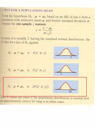
Use of ‘uncorrected’ p-value, a=0.1 11.3% 11.3% 12.5% 10.8% 11.5% 10.0% 10.7% 11.2% 10.2% 9.5% Percentage of Null Pixels that are False Positives Using an ‘uncorrected’ p-value of 0.1 will lead us to conclude on average that 10% of voxels are active when they are not. This is clearly undesirable. To correct for this we can define a null hypothesis for images of statistics.
FAMILY-WISE NULL HYPOTHESIS: Activation is zero everywhere If we reject a voxel null hypothesis at any voxel, we reject the family-wise Null hypothesis A FP anywhere in the image gives a Family Wise Error (FWE) Family-wise Null Hypothesis Family-Wise Error (FWE) rate = ‘corrected’ p-value
Use of ‘uncorrected’ p-value, a=0.1 Use of ‘corrected’ p-value, a=0.1 FWE
Spatial correlation Independent Voxels Spatially Correlated Voxels
Consider a statistic image as a discretisation of a continuous underlying random field Use results from continuous random field theory Random Field Theory Discretisation
Topological measure threshold an image at u EC=# blobs at high u: Prob blob = avg (EC) so FWE, a = avg (EC) Euler Characteristic (EC)
Example – 2D Gaussian images • α = R (4 ln 2) (2π) -3/2 u exp (-u2/2) Voxel-wise threshold, u Number of Resolution Elements (RESELS), R N=100x100 voxels, Smoothness FWHM=10, gives R=10x10=100
Example – 2D Gaussian images • α = R (4 ln 2) (2π) -3/2 u exp (-u2/2) For R=100 and α=0.05 RFT gives u=3.8
Outline • Voxel-wise General Linear Models • Random Field Theory • Bayesian Modelling
Motivation Even without applied spatial smoothing, activation maps (and maps of eg. AR coefficients) have spatial structure Contrast AR(1) We can increase the sensitivity of our inferences by smoothing data with Gaussian kernels (SPM2). This is worthwhile, but crude. Can we do better with a spatial model (SPM5) ? Aim: For SPM5 to remove the need for spatial smoothing just as SPM2 removed the need for temporal smoothing
q1 q2 a b u1 u2 l W A Y The Model r2 r1 Voxel-wise AR: Spatial pooled AR: Y=XW+E [TxN] [TxK] [KxN] [TxN]
Synthetic Data 1 : from Laplacian Prior reshape(w1,32,32) t
Prior, Likelihood and Posterior In the prior, W factorises over k and A factorises over p: The likelihood factorises over n: The posterior over W therefore does’nt factor over k or n. It is a Gaussian with an NK-by-NK full covariance matrix. This is unwieldy to even store, let alone invert ! So exact inference is intractable.
L KL F Variational Bayes
If you assume posterior factorises then F can be maximised by letting where Variational Bayes
Variational Bayes In the prior, W factorises over k and A factorises over p: In chosen approximate posterior, W and A factorise over n: So, in the posterior for W we only have to store and invert N K-by-K covariance matrices.
Observation noise Updating approximate posterior Regression coefficients, W AR coefficients, A Spatial precisions for A Spatial precisions for W
F Iteration Number
VB – Laplacian Prior Least Squares y y x x Coefficients = 1024 `Coefficient RESELS’ = 366
Synthetic Data II : blobs Smoothing True Global prior Laplacian prior
Sensitivity 1-Specificity
Event-related fMRI: Faces versus chequerboard Smoothing Global prior Laplacian Prior
Event-related fMRI: Familiar faces versus unfamiliar faces Smoothing Penny WD, Trujillo-Barreto NJ, Friston KJ. Bayesian fMRI time series analysis with spatial priors. Neuroimage. 2005 Jan 15;24(2):350-62. Global prior Laplacian Prior
Summary • Voxel-wise General Linear Models • Random Field Theory • Bayesian Modelling http://www.fil.ion.ucl.ac.uk/~wpenny/mbi/index.html Graph-partitioned spatial priors for functional magnetic resonance images. Harrison LM, Penny W, Flandin G, Ruff CC, Weiskopf N, Friston KJ. Neuroimage. 2008 Dec;43(4):694-707.