Levels of Protein Structure
Levels of Protein Structure. Primary to Quaternary Structure. Learning Objectives. After today you should be able to: Define the structural levels of proteins. Identify the structural units of the protein backbone.

Levels of Protein Structure
E N D
Presentation Transcript
Levels of Protein Structure Primary to Quaternary Structure
Learning Objectives • After today you should be able to: • Define the structural levels of proteins. • Identify the structural units of the protein backbone. • Explain why some backbone conformations are “forbidden”, i.e. not found in natural proteins. • Name properties on which the amino acids can be grouped. • Name more amino acids than you could before One and three letter codes
Proteins are built from amino acids Amino group and acid group Side chain at Ca Chiral, only one enantiomer found in proteins (L-amino acids) 20 natural amino acids Amino Acids Ce Sd Cg Cb C Ca N O Methionine
L-amino acids in Living organisms N: 14 C: 12 O: 16
How to Group Them? Many features Charge +/- Acidic vs. basic (pKa) Polarity (polar/non-polar) Type, distribution Size Length, weight, volume, surface area Type (Aromatic/aliphatic)
The Amino Acids Thr (T) Phe (F) Val (V) Ala (A) His (H) Arg (R) Ser (S) Leu (L) Cys (C) Met (M) Asp (D) Lys (K) Asn (N) Ile (I) Trp (W) Gln (Q) Glu (E) Tyr (Y) Pro (P) Gly (G)
A – Ala C – Cys D – Asp E – Glu F – Phe G – Gly H – His I – Ile K – Lys L – Leu M – Met N – Asn P – Pro Q – Gln R – Arg S – Ser T – Thr V – Val W – Trp Y - Tyr Amino Acids Livingstone & Barton, CABIOS, 9, 745-756, 1993
The Evolution Way • Based on Blosum62 matrix • Measure of evolutionary substitution probability
The Simple Aliphatic Val (V) Ala (A) Leu (L) Met (M) Ile (I)
The Small Polar Ser (S) Cys (C) Thr (T) Cystin
The Unusual • P, G • Also aliphatic • Structural impact • Strictly speaking, proline is an imino acid Gly (G) Pro (P)
The Acidic and Their Derivatives Asp (D) Asn (N) Glu (E) Gln (Q)
The Basic Arg (R) Lys (K) His (H)
The Aromatic Phe (F) His (H) Trp (W) Tyr (Y)
The peptide bond A polypeptide chain Proteins Are Polypeptides
N H Ramachandran Plot • Allowed backbone torsion angles in proteins
For the polypeptide on the left discuss the following with your neighbour: Why is the lower right quadrant a ”forbidden” region in the Ramachandran plot? What makes Gly a special amino acid when it comes to Ramachandran plots? What about Pro? Small Exercise (5 minutes)
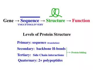
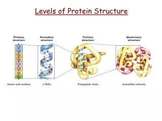
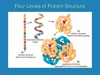
Primary structure = Sequence (of amino acids) Secondary Structure = Helix, sheets/strands, bends, loops & turns (all defined by H-bond pattern in backbone) Structural Motif = Small, recurrent arrangement of secondary structure, e.g. Helix-loop-helix Beta hairpins EF hand (calcium binding motif) Many others… Tertiary structure = Arrangement of Secondary structure elements within one protein chain Structure Levels MSSVLLGHIKKLEMGHS…
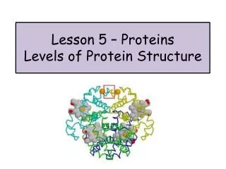
Myoglobin Haemoglobin Assembly of monomers/subunits into protein complex Backbone-backbone, backbone-side-chain & side-chain-side-chain interactions: Intramolecular vs. intermolecular contacts. For ligand binding side chains may or may not contribute. For the latter, mutations have little effect. a a b b a Quaternary Structure
Hydrophobic Core • Hydrophobic side chains go into the core of the molecule – but the main chain is highly polar. • The polar groups (C=O and NH) are neutralized through formation of H-bonds. Myoglobin Surface Interior
Globular protein (in solution) Membrane protein (in membrane) Hydrophobic vs. Hydrophilic Myoglobin Aquaporin
Globular protein (in solution) Membrane protein (in membrane) Hydrophobic vs. Hydrophilic Cross-section Cross-section Myoglobin Aquaporin
Characteristics of Helices • Aligned peptide units Dipolar moment • Ion/ligand binding • Secondary and quaternary structure packing • Capping residues • The a helix (i→i+4) • Other helix types! (310, p) C N
b-Sheets • Multiple strands sheet • Parallel vs. antiparallel • Twist • Coil regions between the secondary structure elements Thioredoxin
b-Sheets • Multiple strands sheet • Parallel vs. antiparallel • Twist
b-Sheets • Multiple strands sheet • Parallel vs. antiparallel • Twist
b-Sheets • Multiple strands sheet • Parallel vs. antiparallel • Twist
b-Sheets • Multiple strands sheet • Parallel vs. antiparallel • Twist • Strand interactions are non-local • Flexibility • Vs. helices • Folding Antiparallel Parallel
Summary • Amino acids in Living organisms have L-configuration • One & three letter codes • Groups of amino acids: hydrophobic ... • The backbone of polypeptides form regular secondary structures. • Helices, sheets & loops.
Building Blocks Ca Peptide unit (amide) COOH OH O C N C/CH/CH2/CH3 O Amide C Indole N S/SH H-bond Phenyl Imidazole Guanidine NH2/NH3+
Building Blocks Ce Sd c3 On-1 Nn+1 Cg Ca Ca O C N C N Cn-1 c2 Cb O O c1 Ca y Single amino acid N Residue f C O O Methionine
Build backbone in extended conformation (strand). Twist to align all peptide units (look at the C=O groups). Add H-bonds (i+4) to construct an ideal a-helix. Add side chains along the way (pointing “down”). Procedure C N