Download
1 / 19
260 likes | 1.12k Vues
Central Venous Access Devices (VADs). Management Complications Troubleshooting. Objectives. The learner will be able to: Describe general principles associated with the proper care of each type of VAD. Identify key complications associated with central venous access catheters.

E N D
Central Venous Access Devices(VADs) Management Complications Troubleshooting
Objectives The learner will be able to: • Describe general principles associated with the proper care of each type of VAD. • Identify key complications associated with central venous access catheters.
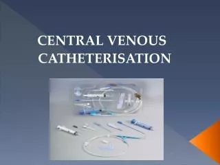
General Principles of Care for VADs • Maintain strict aseptic technique for all procedures. • Wear appropriate PPE for all procedures. • Secure all tubing connections with Luer locks. • Procedures should be performed by personnel who know how to assess for complications. • Avoid using tape to secure connections (may cause bacterial transmission). • Change dressing, IV tubing, or protective cap if wet, soiled, contaminated, or damaged. • Includes nontunneled, PICC, tunneled, and implanted port catheters.
General Principles of Care: VAD Dressing Changes • Change 24 hours after insertion. • Change transparent dressing every 57 days. • Change gauze dressing QOD or PRN if wet/soiled. • Tunneled: No dressing once healed unless immunocompromised • Implanted port: For continuous access, change noncoring needle/transparent dressing every week; gauze QOD or PRN if wet/soiled.
General Principles of Care:VAD Flushing Procedures • Essential to ensure patency and prevent accumulation of blood/drug precipitates in the catheter lumen • Can prevent fibrin buildup but all VADs will accumulate fibrin coating to some extent • Locking solutions used to prevent occlusions when the device is not in use • Never use excessive force when flushing. • Typical flush: Heparin 10100 IU/ml, 3 ml/day or QOD unless in maintenance
Principles of Care: Central VADs or Central Venous Catheters (CVCs) • Verify catheter placement. • Inspect site for erythema, swelling, drainage, and leakage. • Inspect ipsilateral (same side of) chest for signs of venous thrombosis. • Aspirate to verify blood return. • Obtain a physician’s order for declotting, if needed. • Use x-rays or dye studies to confirm placement.
Principles of Care: Accessing and Deaccessing of Implanted Ports • Must be accessed with noncoring needle • When not used, must be accessed and flushed every 46 weeks • Positive pressure technique is extremely important to prevent fibrin sheath development.
Principles of Care: Implanted Ports • Wear appropriate PPE. • Assess initial line placement and for signs of infection. • Use a noncoringneedle. • Prepare patient’s skin. • Access the port. • Establish blood return and patency. • Inspect the needle insertion site for needle dislodgement, leakage of fluid, and edema. • Examine ipsilateral chest for signs/symptoms of venous thrombosis. • Apply occlusive dressing to stabilize the needle.
Complications: CVCs • CVCs should be used only when the benefit outweighs the risks. • Except for a pulmonary artery catheter, the tip should not be placed in or allowed to migrate into the heart. The distal tip should be in the superior vena cava at the junction of the right atrium. • Tip location should be confirmed by x-ray or other imaging; recheck if not functioning or no blood return. • Catheters placed in less-than-sterile environments should be replaced as soon as possible.
Complications: CVCs:Post-Insertion Catheter Migration • Signs/symptoms • Ability to infuse changes/unable to withdraw blood • Increased length of external catheter • Arm/shoulder pain • Swelling • Chest pain • Sensation of “gurgling” in the throat • Arrhythmia • Intervention • Confirm placement by x-ray or cathetergram.
Complications: CVCsPinch-Off Syndrome • The anatomic, mechanical compression of the catheter between the clavicle and first rib at the costoclavicular space • Signs/symptoms • Difficulty infusing or withdrawing blood or able to do so with patient repositioning • Symptoms of fracture: Arrhythmia, extra heart sounds, extravasation, shortness of breath, palpitations • Intervention • Chest x-ray and if confirmed, must be removed
Complications: CVCsOcclusions • Etiologies • Intraluminal blood/fibrin, precipitates/lipid deposits, fibrin tail or sheath, mural thrombosis, improperly inserted port needle • Signs/symptoms • Inability to withdraw blood but able to infuse, or inability to withdraw and to infuse, or difficulty unless patient is repositioned • Pain or edema in neck (indicative of a thrombus) • Intervention • Flush catheters well as preventive measure. • Other interventions have their own risks and are dependent upon etiology of occlusion.
Complications: CVCsInfection • Local: At insertion site or port pocket or tunnel • Systemic: Serious complication (septicemia) • Etiologies • Insertion contamination • Neutropenic patient at time of insertion • Contaminated infusate • Seeding from another infectious source • Immunocompromised patient • Hub contamination
Complications: CVCsInfection (cont) • Prevention • Frequent hand washing! • Strict maintenance of aseptic technique • Proper site selection • Daily review of line necessity • Chlorhexidine skin antisepsis • Alcohol hub decontamination before hub access • Signs/symptoms • Local: Swelling, tenderness, erythema, induration, cellulitis, drainage • Systemic: Fever, chills, fatigue, diaphoresis, hypotension, weakness, tachycardia, mental changes, abdominal pain, hyperventilation, vomiting, diarrhea
Complications: CVCsInfection (cont) • Interventions • Assess daily. • Local: Culture location. Apply warm compresses. • Give IV or oral antibiotics as ordered. • Systemic: Draw blood cultures from device and from peripheral blood. Culture infusate. Give IV antibiotics. • If symptoms persist 2448 hours after antibiotics are initiated, remove VAD.
Complications: CVCsDamage • Etiologies: Dislodgement, sheared or fractured by forceful flushing, cut, skin integrity loss over port • Signs/symptoms: Leaks, wet dressings, pain, edema, visualization of port body • Intervention: X-ray or cathetergram for verification; remove if catheter separated from port body or internally damaged • Repair possible if implanted port (2-pieces) and can replace damaged section
Complications: CVCsExtravasation • Causes • Incomplete port needle insertion/dislodged needle • Separation of port body from catheter • Inadequately secured catheter • Catheter tip migration • Prevention • Appropriate needle length to ensure septum penetration • Adequate needle stabilization/use of noncoring needle • Frequent blood return check during vesicant administration: Never give vesicants without a blood return. • Frequent site assessment • Teach patient to report signs and symptoms immediately.
Complications: CVCsExtravasation (cont) • Symptoms • Redness, edema, pain, burning, lack of blood return, leaking, difficulty infusing • Intervention • Stop infusion, aspirate residual drug, assess, give antidote if indicated, apply cold or heat as appropriate. • Notify physician, observe, and document.
References Camp-Sorrell, D. (Ed.). (2011). Access device guidelines: Recommendations for nursing practice and education (3rd ed.). Pittsburgh, PA: Oncology Nursing Society. Polovich, M., Whitford, J.M., & Olsen, M. (Eds.). (2009). Chemotherapy and biotherapy guidelines and recommendations for practice (3rd ed.). Pittsburgh, PA: Oncology Nursing Society. Smith, L. (2008). Implanted ports, computed tomography, power injectors, and catheter rupture. Clinical Journal of Oncology Nursing, 12, 809812.