CENTRAL VENOUS CATHETERISATION
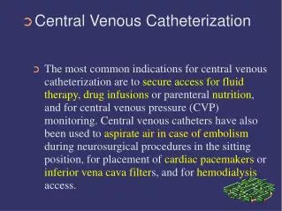
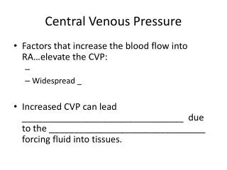
CENTRAL VENOUS CATHETERISATION. INDICATIONS. Measurement of central venous pressure (CVP) ( E.G. those with hypotension not responding to normal management, requiring infusion of inotropes) For long term administration of drugs for pain, infection, cancer or to supply nutrition.

CENTRAL VENOUS CATHETERISATION
E N D
Presentation Transcript
CENTRAL VENOUS CATHETERISATION
INDICATIONS • Measurement of central venous pressure (CVP) ( E.G. those with hypotension not responding to normal management, requiring infusion of inotropes) • For long term administration of drugs for pain, infection, cancer or to supply nutrition. • Venous access for IV fluids or antibiotics or a peripheral site is unavailable/unaccessible • Haemodialysis
CONTRAINDICATIONS • Patients undergoing thrombolytic or anticoagulative therapy • Bleeding disorders • Vasculitis • Distorted local anatomy • Overlying skin infections( dermatitis), burns • Uncooperative patient
COMPLICATIONS • INFECTIOUS Sepsis (also septic arthritis, osteomyelitis) • VASCULAR Air embolism, blood clot, hematoma, arterial puncture • OTHERS PNEUMOTHORAX, hemothorax, arrhythmias , nerve injury
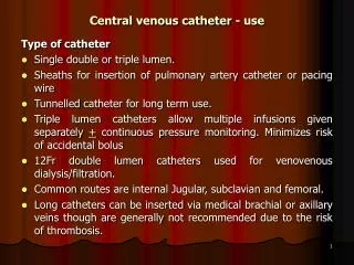
LOCATIONS of CVC • INTERNAL JUGULAR VEIN • SUBCLAVIAN VEIN • FEMORAL VEIN Length of catheters • 15cm catheters for subclavian and internal jugular lines, and 60cm catheters for femoral line
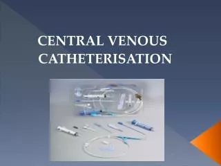
EQUIPMENT USED: • Patient on a tilting bed, trolley or operating table • Standard multiple lumen kit • Guide wire • Sterile gloves • Sterile gown • Drapes • Disinfectant (Povidone-iodine solution/ chlorhexidine) • Suturing needle • Scalpel • Local Anaesthetic (lidocaine) • Sterile saline flush
Triple lumen catheter Dilator A= small syringe and vial of 1% Lidocaine (L.A) B= guide needle C= IV syringe with catheter attached- D E= Disinfectant sponge Guidewire: J-shaped tip to reduce risk of vessel perforation
TECHNIQUE • SELDINGER TECHNIQUE (most common) • Use guide needle to locate the vein • Wire threaded through needle • Remove needle • A dilator is passed over the guide wire • Dilator is removed and catheter is passed over wire and wire is removed • Catheter secured in place Allowslarger catheters to be placed in the vein after the passage of appropriate dilators along the wire and a small incision in the skin at the point of entry.
GENERAL PRINCIPLES • Obtain informed consent and explain risks and benefits of procedure • Optimal patient positioning and cooperation, make sure patient is comfortable • Take your time • Sterile technique • Local anaesthetic should be used • Always have a hand on your wire • Aspirate while advancing as you withdraw the needle slowly • Withdraw needle to the level of the skin before redirecting the angle • Don’t poke yourself with the needle • The tip of the catheter can lie in either the superior or inferior vena cava (SVC or IVC) or into the right atrium (RA).
INTERNAL JUGULAR APPROACH • POSTIONING : TRENDELENBURG POSITION • Patient supine on surface inclined 45 degrees, head at the lower end and legs flexed over upper end. ( This distends the central veins and prevents air embolism) • Head turned to opposite side of central venous line • Stand at the head of the patient
LOCATING AND ACCESSING VEIN • Ultrasound and landmarks can be used • IJV is between the clavicular and sternal heads of the sternocleidomastiod muscle • Point of needle insertion is midway between sternal head of SCM and mastoid process behind ear
PROCEDURE • Disinfect area , apply L.A and fenestrated drape • Place three fingers on carotid artery • Place needle about 45 degrees to the skin, lateral to the carotid artery • Direct needle in sagittal plane angled towards feet • Vein should be 1-1.5 cm deep, avoid deep probing in the neck • Seldinger technique used http://www.youtube.com/watch?v=QHiuYc22pfE
2. Insert needle into IJV and aspirate 3. Hold tip of needle with one hand 1.Disinfection, L.A and sterile drape 6. Once catheter is in place , secure and apply dressing 4. Place wire through needle and remove needle 5. Insert catheter over wire then remove wire
SUBCLAVIAN APPROACH POSITIONING • Trendelenburg postion (10-15 degrees) • Supine position, head and shoulders neutral with arm slightly abducted • Stand beside the patient at the side PROCEDURE • Disinfect area and apply local anaesthetic • Cover procedure area with fenestrated sterile drape • Use seldinger technique LOCATION AND ACCESS TO VEIN • Identify the midclavicular point and sternal notch • Insert needle into the skin 1cm below and lateral to the midclavicular point • Direction of needle should be parallel to skin • Advance posterior to the clavicle aiming for the sternal notch (Do not pass the needle further than the sternal head of the clavicle)
Location and insertion of needle http://video.google.com/videoplay?docid=2421324986948418582#
FEMORAL VEIN APPROACH POSITIONING • Supine patient • Extend the patient’s leg and abduct slightly at the hip PROCEDURE • Disinfect area and apply local anaesthetic • Cover procedure area with fenestrated sterile drape • Use seldinger technique LOCALISATION AND ACCESS TO VEIN • Vein is medial to femoral artery • Identify the pulsation of the femoral artery 1-2 cm below the inguinal ligament. • Position needle at 45 degree angle and about 1cm medial to pulsation • Needle inserted at skin about 2 cm below inguinal ligament • Aiming towards the umbilicus (In adults, the vein normally found 2-4cm from the skin. In small children reduce elevation on the needle to 10-15° as the vein is more superficial)
http://www.4shared.com/file/61538831/b1027127/Placement_of_a_Femoral_Venous_.htmlhttp://www.4shared.com/file/61538831/b1027127/Placement_of_a_Femoral_Venous_.html
POST CATHETER PLACEMENT • Aspirate blood from each port • Flush with saline / sterile water • Secure the catheter with sutures • Apply sterile dressing • Dispose of used gloves, needles, syringe etc • Wash hands • Chest x-ray for IJ and SC lines