Download
1 / 32
640 likes | 2.41k Vues
Central venous catheters. Kristin wise, md, fhm. Objectives. Understand the indications & contraindications for Central Venous Catheters (CVCs) Identify factors that influence the selection of appropriate site for CVC insertion Describe the procedural steps for CVC placement

E N D
Central venous catheters Kristin wise, md, fhm
Objectives • Understand the indications & contraindications for Central Venous Catheters (CVCs) • Identify factors that influence the selection of appropriate site for CVC insertion • Describe the procedural steps for CVC placement • Recognize common CVC complications
Key messages • CVC indications include medication administration, hemodynamic monitoring, poor peripheral IV access & venous access to place other devices. • There are no absolute contraindications for a CVC. Relative contraindications depend on patient specific factors. • While the preferred CVC insertion site varies between patients, subclavian (SC) & internal jugular (IJ) veins have significantly fewer infectious & mechanical complications than femoral veins. • AHRQ Safety Practices recommend US guidance and use of maximal sterile barriers when inserting CVCs. • The Seldinger technique uses an access needle, guidewire & dilator to introduce CVCs safely into vessels. • Common immediate complications include arterial puncture, hematoma & PTX. Common delayed complications include infection & thrombosis.
overview • Described first in 1952 by Dr. Aubaniac • Cannulatedsubclavian vein in battlefield to resuscitate wounded soldiers • ACGME, ABIM, SHM recognize CVCs as a ‘competency’ for internal medicine physicians • ~8% hospitalized pts require CVCs • >5 mil CVCs inserted annually in US • >15% pts have complications
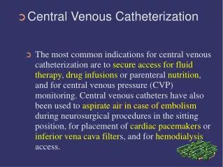
Indications for CVCS • Administer Meds or Nutrition • Infuse meds (ie, vasopressors, chemotherapy) or total parenteral nutrition (TPN) that cause phlebitis/sclerosis when given through peripheral IVs • Hemodynamic monitoring • Measurement of central venous pressure, venous oxyhemoglobin saturation & cardiac parameters (via pulmonary artery catheter). • Poor peripheral venous access • Perform ultrafiltration, plasmapheresis/apheresis, HD or other blood filtering process • Provide venous sheath access to place other devices • IVC filters, venous stents, cardiac pacemakers/defibrillators, etc.
contraindications No absolute contraindications for CVC placement Important relative contraindications: • Coagulopathy • target values: INR <1.5, PTT <40 • Thrombocytopenia • target value: Plts >50K • No literature to support correcting these abnormalities • Expert opinion recommends most experienced individual available perform procedure • Avoid subclavianvein • Difficult to monitor bleeding & effectively compress venipuncture site
relative contraindications • Infected area overlying target vein • Anatomic distortion or injury proximal to insertion site • Other indwelling intravascular hardware (ie, pacemaker, hemodialysis catheter) • Fracture or suspected fracture of clavicle or proximal ribs (for subclavian) • Thrombosis of target vein • Complications would be life-threatening (ie, PTX in hypoxic pt)
Site selection PROs CONs
Site selection: other considerations • Multiple scars from prior access • Unilateral lung disease (ie, PNA, traumatic PTX) • Place access ON disease side to minimize respdecompensationfrom procedure-related PTX • Future need for HD • Avoid subclavian due to risk of catheter-induced vein stenosis which complicates/eliminates subsequent HD access options • Cerebral venous outflow in acute stroke or neuropts • Subclavian site preferred over IJ
Site selection: considering complications Remember these complications to obtain informed consent
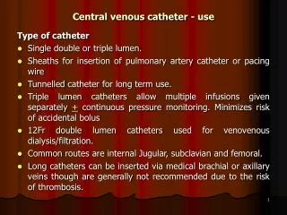
Catheter descriptions Types: • Central • Peripheral (think PICC line) • Introducer sheaths (think pul artery catheter) Size: • Diameter: 7-French most common 8.5-French common for introducers 11.5-French common for HD • Length: 15cm to 30cm for central lines <10cm for introducers
Catheter descriptions • Most polyurethane or silicone • Specialized catheters exist-–valve mechanisms, power-injectable, antibiotic-impregnated or heparin-coated Lumens—single, double, triple, quadruple • ↑ number of lumens means ↑ CVC diameter • More lumens does NOT mean a better catheter • ↓ individual lumen diameter thus ↓ max infusion rates • ↑ rate of catheter thrombosis • correlates with ↑ mechanical complications
Catheter descriptions Lumens— • infuse fluid through end & holes located on side of catheter • end hole more reliable for blood draws (less likely suctioned against vein wall)
Preparation for insertion • Review labs or imaging • Use clinical info to determine best site • US site to confirm vessel patency • Review allergies to meds & tape • Obtain informed consent • Position patient for procedure AND your comfort • Supine, Trendelenberg15-30◦ • Rotate head 45◦ away from cannulationsite • Monitor if possible (telemetry, pulsox) • O2 available, consider O2 use prior to draping patient • ‘Universal Time-Out’
Safety practices AHRQ (2001)—Top 11 most highly-rated safety practices • List based on strength of evidence to support widespread implementation “Use of real-time US guidance during CVC insertion to prevent complications” “Use of maximum sterile barriers while placing CVCs to prevent infections”
Ultrasound guidance • High frequency probe (5-10MHz) • Scan potential insertion sites • Evaluate venous patency with compression • Facilitate patient positioning to optimize vessel location • Identify vein • Non-pulsatile, compressible (if hypotensive, artery may compress) • Often irregularly shaped compared to artery • Thinner vessel wall than artery • Often bigger than artery (if volume depleted, may be smaller) • ↓ time to venous cannulation • ↓cannulation attempts, ↓ complications
Sterile technique • Non-sterile mask & cap for everyone present • Non-sterile cap on patient • Proper hand hygiene • Wear sterile gown & gloves • Open sterile kit & get organized • Prepare/flush all ports of CVC • Prep site • 30sec continuous scrubbing with Chlorhexidine • allow site to fully dry • Use wide sterile drape • Use sterile US probe cover
IJ vein Landmarks: • apex of triangle formed by SCM heads & clavicle • lateral to carotid pulse • aim for ipsilateral nipple
SC vein Landmarks: • crosses under clavicle just medial to mid-clavicular point • ~2cm lateral, ~2cm caudal to middle third of clavicle
Procedure steps • Re-identify vein on US • Use non-dominant hand to hold US probe • Anesthetize cannulation site • Use 1% lidocaine with 25g needle • Aspirate before injecting (lidocaine causes vasospasm & systemic effects!) • Only use 1-2 ml to avoid creation of distracting fluid pocket on US
Seldinger technique Dr. Sven-Ivar Seldinger (1921-1998) Swedish radiologist Technique introduced In 1953 to obtain safe access to blood vessels & hollow organs Modified version in use today.
Seldinger technique Cannulate vein with US guidance (preferred) • Advance needle at 45◦ angle • Maintain negative pressure (aspirate) on syringe as advance needle until vein punctured Use non-dominant hand (set aside US) to grasp needle • Brace hand against pt’s body to stabilize & maintain needle position • Lower angle of needle Disconnect syringe with dominant hand • If no blood seen from hub: Reconnect immediately Aspirate to ensure proper needle placement Consider ↑ Trendelenburg
Seldinger technique • Insert guidewire through needle • Should thread easily, w/o resistance, don’t force it! • Never let go of wire • Don’t over-insert guidewire • End of guidewire remains longer than pt’s head (IJ) or shoulder (SC) • If arrhythmia noted, pull wire back until rhythm normalizes • Remove needle while controlling guidewire • Never let go of wire • Use scalpel to make skin nick adjacent to wire
Seldinger technique • Advance dilator over guidewire • Don’t over-insert dilator • Advance it ≤ 5cm from skin surface • Non-obese, shallow vessel only insert ~2cm • Never let go of wire • Remove the dilator • Never let go of wire
Seldinger technique • Thread catheter over guidewire • Hold wire taut to make catheter slide on wire easier • Don’t accidentally pull wire back until catheter in • Never let go of wire • Catheter indwelling length depends on insertion site & pt size • Average guidelines: • RIJ 13cm, LIJ 18-20cm • RSC 15cm, LSC 18cm • Femoral 20cm or longer
Seldinger technique • Remove guidewire • Now you can safely let go of wire • Aspirate blood from all ports & flush with saline • Secure catheter with sutures • Do not overtightensutures, can lead to skin necrosis then sutures come out • Apply sterile dressing • Use Biopatch if available, “blue” side points up to “sky” • Remove sterile drape carefully • Do not dislodge CVC, it can get caught in drape
Post-procedure etiquette • Obtain CXR (IJ, SC) to confirm catheter tip position • Tip in right atrium or SVC • Malposition of tip increases venous thrombosis risk • CXR also allows evaluation for PTX • Clarify with RNs whether line ready to use or if CXR required • OK to draw urgent/emergent labs from functioning line w/o CXR results • Clarify line flushing orders—saline or heparin? • Write procedure note
Procedure note • Name of procedure performed • Date & time • Indication • Clinician(s) involved • Refer to Informed Consent & Universal Time-Out • Note use of wide sterile barriers & proper hand hygiene • Was US used? • Vessel(s) cannulated, number of attempts • Type catheter, # lumens, insertion length • Patient status during & after procedure • Whether post-procedure CXR ordered • Relevant findings/events during procedure
Review of Complications Most common: • Arterial puncture • Hematoma • PTX Most common: • Infection • DVT
Complication rates • Incidence rate of complications 6x higher with ≥3 insertion attempts vs. single attempt • US can improve 1st attempt success • Operator experience: Mechanical complications 50% higher with MD who has placed ≤50 CVCs vs. MD who has placed ≥50 CVCs • Most experienced person available to place CVC on complicated, unstable pts
Key references Rosen BT, Wittnebel K. Central line placement. In: McKean SC, Ross JJ, Dressler DD, Brotman DJ, Ginsberg JS, eds. Principles and Practice of Hospital Medicine. 1st ed. New York, NY: McGraw-Hill; 2012:860-65. McGee DC, Gould MK. Preventing complications of central venous catheterization. NEJM. 2003;348(12):1123-33. Taylor RW, Palagiri AV. Central venous catheterization. Crit Care Med. 2007;35(5):1390-96. Web-based resources Heffner AC, Androes MP. (2013). Overview of central venous access. In: Collins KA, ed. UpToDate. Available from http://www.uptodateonline.com. Web-based resources: Videos in Clinical Medicine Graham AS, OzmentC, TegtmeyerK, Lai S, BranerDV. Central venous catherization. NEJM. 2007;356:e21. Braner DAV, Lai S, EmanS, Tegtmeyer K. Central venous catherization—Subclavian vein. NEJM. 2007;357:e26.