Exploring the Brain: Vision and Seizures
180 likes | 208 Vues
Delve into the fascinating world of brain functions, epilepsy, and seizure disorders. Learn about the split-brain discoveries, causes of seizures, epilepsy treatments, and innovative brain study methods.

Exploring the Brain: Vision and Seizures
E N D
Presentation Transcript
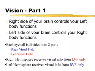
Vision - Part 1 • Right side of your brain controls your Left body functions • Left side of your brain controls your Right body functions • Each eyeball is divided into 2 parts • Right Visual Field • Left Visual Field • Right Hemisphere receives visual info from LVFonly • Left Hemisphere receives visual info from RVFonly
Vision - Part 2 • Left Visual Field is illustrated in RED • Right Visual Field is illustrated in BLUE Lesson Outline Split-Brain Discoveries
Ways to study the Brain!!! Accidents: damage to brain regions can tell us about their functions Phineas Gage.
Lesions: tissue destruction Cutting into the brain and looking for change. Brain tumors also lesion brain tissue.
Seizures • 3 basic types • Grand Mal • Involves total body convulsions, aka “tonic-clonic” • Petit Mal • Involves isolated body part convulsion, aka “focal” • Absence • Patient becomes unresponsive, and has no memory of occurrence. Appears to be day-dreaming but cannot awake. Very rare.
What is a seizure? 1. Abnormal discharge of electrical impulses within the brain 2. Rather than smooth constant production of Action Potentials, neurons fire without any regulation, causing disruption to brain function at the biochemical level • Seizures generally have 3 parts: Aura - period of warning, usually olfactory or visual Ictus - actual seizure period Postictal state - time where body “resets” itself
Causes of Seizures Alcohol Poisoning Brain Tumor Drug Overdose/Reaction Stroke Epilepsy Head Injury Fever (especially in children) Neurological Defect (usually genetic) Sepsis (in brain) Lesson Outline
Epilepsy • A seizure disorder in which reoccurring seizures are the main symptom caused by an abnormal discharge of electrical activity from the neurons in the cerebral cortex. • In the US more than 4 million people have some form of epilepsy • Risk of epilepsy is greatest in early childhood and late adulthood. • Seizures have been found depicted as early as in cavepaintings! • 4,000 yr. old writings depict epileptics as “possessed by demons” • Julius Caesar, King Charles II, Vincent Van Gogh and novelist Dostoyevsky all reportedly suffered from seizures.
Treatments for Epilepsy • 3 major courses of treatment: • Drugs • Generally first line of attack because it is effective, relatively inexpensive, and safe • Diet • Ketogenic diet - lots of fat and almost no carbohydrates • This diet drastically alters the way our bodies get energy from food - instead of making glucose, it makes ketones • Surgery • Commissurotomy
Commissurotomy • For patients with frequent and violent epileptic seizures, surgically splitting the corpus callosum was the only relief - known as a “commissurotomy” • Corpus callosum is a bundle of nerve fibers which serve to connect the right and left cerebral hemispheres
Less Invasive ways to study the Brain Electroencephalogram (EEG) Computerized Axial Tomography (CAT) Positron Emission Tomography (PET) Magnetic Resonance Imaging (MRI) Functional MRI
Electroencephalogram (EEG) • Electrodes placed on the scalp create an amplified recording of the waves of electrical activity that sweep across the brain’s surface
CT scan • CT (computed tomography) Scan • a series of x-ray photographs taken from different angles and combined by computer into a composite representation of a slice through the body; also called CAT scan
PET Scan • PET (positron emission tomography) Scan • a visual display of brain activity that detects where a radioactive form of glucose goes while the brain performs a given task
MRI Scan • MRI (magnetic resonance imaging) • a technique that uses magnetic fields and radio waves to produce computer-generated images that distinguish among different types of soft tissue; allows us to see structures within the brain MRI scan of a healthy individual (left) and a person with schizophrenia (right) Note the enlarged fluid - filled brain region in the image on the right.
fMRI Scan • Functional MRI • Reveals blood flow, and therefore, brain activity by comparing successive MRI scans. • “Reading Your Mind” – 60 Minutes
Brain Restoration – Plasticity • The ability for our brains to form new connections after the neurons are damaged. • Evidenced by brain reorganization following damage (especially in children) and in experiments on the effects of experience on brain development • The younger you are, the more plastic your brain is. • Plasticity of sensory cortex
Brain Restoration Glial Cells • cells in the nervous system that support, nourish, and protect neurons