CTSA at RSNA 2010
860 likes | 1.02k Vues
An end-user application for image analysis and visualization An open-source environment for software development A software platform that is both easy to use for clinical researchers and easy to extend for programmers. CTSA at RSNA 2010. --. www.slicer.org. Disclaimer

CTSA at RSNA 2010
E N D
Presentation Transcript
An end-user application for image analysis and visualization • An open-source environment for software development • A software platform that is both easy to use for clinical researchers and easy to extend for programmers CTSA at RSNA 2010 --
www.slicer.org Disclaimer It is the responsibility of the user of 3DSlicer to comply with both the terms of the license and with the applicable laws, regulations and rules. Slicer is a tool for research, and is not FDA approved. 3D Slicer version 3 is a multi-platform software running on Windows, Linux, and Mac OSX. CTSA at RSNA 2010 --
This workshop uses the newest release of 3D Slicer (version 3.6.2). • Visit the Slicer download page for Slicer 3.6 stable release, or for Slicer nightly builds. CTSA at RSNA 2010 --
Tutorial Overview • Getting Started: Slicer3 Minute Tutorial • Quantitative Measurement of Volumetric Change: ChangeTracker Tutorial • Quantitative Measurements for Functional Imaging: PETCTFusion Tutorial All Tutorial Datasets are located in C:\slicer_data CTSA at RSNA 2010 --
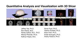
Slicer3 Minute Tutorial Part I: Slicer3 Minute Tutorial Sonia Pujol, PhD Wendy Plesniak, PhD This tutorial is a short introduction to the advanced 3D visualization capabilities of the Slicer3 software for medical image analysis. It is designed to quickly build a basic level of comfort with the Slicer software. CTSA at RSNA 2010 --
Slicer3 Minute Tutorial The Slicer3Minute dataset is composed of An MR scan of the brain and 3D surface Reconstructions of anatomical structures. The data are part of the SPL-PNL Brain Atlas Developed by Talos et al. The atlas is available at: http://www.spl.harvard.edu/publications/item/view/1265 CTSA at RSNA 2010 --
Slicer3 Minute Tutorial: Launch the Application Windows users: Double-Click the Shortcut to Slicer3.exe on the Desktop or Select Start ->All Programs ->Slicer3 3.5.2009-11-06->Slicer CTSA at RSNA 2010 --
Slicer3 Minute Tutorial: Navigating the Application GUI Menu & Toolbar • The Graphical User Interface (GUI) of Slicer3 integrates five components: • the Menu Toolbar • the Module GUI Panel • the 3D Viewer • the Slice Viewer • the Slice and 3D View Controller Module GUI Panel 3D Viewer Slice Viewers Slice and 3D View Controller CTSA at RSNA 2010 --
Slicer3 Minute Tutorial: Welcome Module The SlicerWelcome module is the module displayed by default. This module gives an overview of the GUI of Slicer3, and data loading & saving functionalities. CTSA at RSNA 2010 --
Slicer3 Minute Tutorial: GUI Basics Expand or shrink the GUI panel with the arrows at the frame top, or by clicking and dragging the vertical separator Expand or collapse any sub-panel by clicking on its grey title bar. CTSA at RSNA 2010 --
Slicer3 Minute Tutorial: Load A Scene Select File-> Load Scene from the File menu Browse to the location of the Slicer_data directory. Select that directory and select the Add To Favorites icon This will make the directory easier to find later... CTSA at RSNA 2010 --
Slicer3 Minute Tutorial: Load A Scene Browse to the location of the Slicer3MinuteDataset directory. Select the scene file slicer3minute.mrml Click on Open to load the scene CTSA at RSNA 2010 --
Slicer3 Minute Tutorial: Viewing the Scene 3D Viewer • When the scene is finished loading, Slicer displays: • a 3D model of the head in the 3D Viewer, and • anatomical MR slices of the brain in the 2D Slice Viewers. 2D Slice Viewers CTSA at RSNA 2010 --
Slicer3 Minute Tutorial: Viewing the Scene Note: We have pre-adjusted the window and level settings for these volumes so that they are appropriate for display on most laptops. If display is not satisfactory on your machine or projector, the Volumes Module may be used to refine these settings. CTSA at RSNA 2010 --
Slicer3 Minute Tutorial: Exploring Slicer’s functionality Left click and hold the Modules menubutton. Select All Modules to display the many modules available for image processing, analysis and 3D visualization. CTSA at RSNA 2010 --
Slicer3 Minute Tutorial: Exploring Slicer’s functionality To access the Modelsmodule, browse through the list of modules... ...or click on the models icon in the toolbar CTSA at RSNA 2010 --
Slicer3 Minute Tutorial: Switching to the Models Module Slicer displays the GUI of the Models module. CTSA at RSNA 2010 --
Left-click & drag Slicer3 Minute Tutorial: Basic 3D Interaction Position the mouse in the 3D Viewer. Hold down the left mouse button and drag to rotate the model. CTSA at RSNA 2010 --
Slicer3 Minute Tutorial: Viewing Slices in the 3D Viewer Click on the Slice Visibility icon to display the Axial Slice in the 3D Viewer CTSA at RSNA 2010 --
Slicer3 Minute Tutorial: 3D Visualization Slicer adds a view of the Axial slice in the 3D View. CTSA at RSNA 2010 --
Slicer3 Minute Tutorial: 3D Visualization Select the Skin model. Change the opacity of the model from 1.0 to 0.0. CTSA at RSNA 2010 --
Slicer3 Minute Tutorial: 3D Visualization The model of the skull bone and eyeballs become visible through the model of the skin in the 3D viewer. (skin model opacity = 0.5) CTSA at RSNA 2010 --
Slicer3 Minute Tutorial: 3D Visualization The model of the skin becomes invisible in the 3D viewer. (skin model opacity = 0.0) (skull model opacity = 1.0) CTSA at RSNA 2010 --
Slicer3 Minute Tutorial: 3D Visualization Click on the Slice Visibility icon in the Green Slice Viewer to display the Coronal Slice in the 3D Viewer. CTSA at RSNA 2010 --
Slicer3 Minute Tutorial: 3D Visualization Select the 3D model skull_bone.vtk in the Model Hierarchy and turn on the Clipping option. CTSA at RSNA 2010 --
Slicer3 Minute Tutorial: 3D Visualization Browse through the coronal slices to expose the 3D model of the white matter, and the left and right optic nerves. CTSA at RSNA 2010 --
Slicer3 Minute Tutorial: 3D Visualization Select the 3D model “skull_bone” in the Model Hierarchy, and turn off its Visibility CTSA at RSNA 2010 --
Slicer3 Minute Tutorial: 3D Visualization Scroll the Coronal Slices to display the hemispheric white matter model in the context of the image data in the 3D Viewer. CTSA at RSNA 2010 --
Slicer3 Minute Tutorial: 3D Visualization Select the hemispheric white matter model called hemispheric_white_matter.vtk Turn off its visibility. CTSA at RSNA 2010 --
Slicer3 Minute Tutorial: 3D Visualization Slicer displays the optic nerve, optic chiasm and optic tracts overlaid on the MR images of the brain. CTSA at RSNA 2010 --
Slicer3 Minute Tutorial: 3D Visualization: Zoom the view Windows/Linux users: Position the mouse in the 3D Viewer, hold down the right mouse button and move the mouse down to zoom in. Mac users: Position the mouse in the 3D Viewer, hold down the apple button and the mouse button and move the mouse down to zoom in. CTSA at RSNA 2010 --
Slicer3 Minute Tutorial: 3D Visualization Slicer3 displays a closer view of 3D anatomical structures overlaid on 2D MR slices CTSA at RSNA 2010 --
Slicer3 Minute Tutorial: Summary • This tutorial has demonstrated: • Basic description of the Slicer3 Application Interface • How to load a scene containing volumes and models • How to visualize these different datasets together Next, we will use these building blocks to perform image analysis and visualize quantitative results. CTSA at RSNA 2010 --
ChangeTracker: exploring small volumetric changes Part II: Analyzing Small Volumetric Changes using the ChangeTracker Module Kilian M Pohl, PhD Ender Konugolu, PhD Andriy Fedorov, PhD The module described in this tutorial was tested on Axial 3D SPGR T1 post Gadolinium scans (Voxel dimension: 0.94mm x 0.94mm x 1.20mm, FOV: 240mm, Matrix: 256 x 256) CTSA at RSNA 2010 --
ChangeTracker: Conventional measures of tumor response • Conventional anatomic imaging using CT or MRI are often used to evaluate tumor size and shape • Most clinical trials that evaluate new chemotherapeutic drugs use changes in uni-dimensional or bi-dimensional measurements to assess response (e.g. RECIST) • Slicer has several tools for applying RECIST methodologies CTSA at RSNA 2010 --
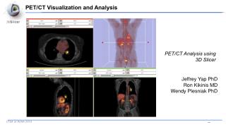
Quantifying tumor change: Conventional measures of tumor response 3D Slicer’s Fiducials Module can be used to measure the longest diameter in a tumor cross section. Two fiducials may be used to mark the tumor’s extent. The distance measurement (mm) between fiducials will be updated in the Fiducial’s GUI. CTSA at RSNA 2010 --
Quantifying tumor change: Conventional measures of tumor response 3D Slicer’s Measurements module, provides interactive measurement tools that operate in the 3D Viewer and the Slice Viewers. Interactive ruler can be used to measure the longest diameterand providesnumericalannotations CTSA at RSNA 2010 --
Quantifying tumor change: Conventional measures of tumor response 3D Slicer’s LabelDiameterEstimation (extension) module will automatically compute the largest tumor diameter and orthogonal dimension. This analysis requires an initial segmentation (VOI). Baseline: June 2006 Follow-up: June 2007 CTSA at RSNA 2010 --
ChangeTracker: rationale for new approaches • However, more accurate and precise methods for understanding volume changes may be useful when: • benign tumor change is being monitored, or • where small changes may be clinically significant but difficult to assess with RECIST • ChangeTracker Module is a tool to measure volumetric change at the voxel level. CTSA at RSNA 2010 --
ChangeTracker: exploring small volumetric changes First, close any previous scene. Select File->Close Scene This removes any datasets previously loaded into Slicer. CTSA at RSNA 2010 --
ChangeTracker: Load the training dataset Select File->Load Scene This raises the Load Scene Interface Select Slicer_data from favorites panel. Select the ChangeTracker2010 directory And select the scene file: ChangetrackerTutorial2009.mrml double click the file, or click Open CTSA at RSNA 2010 --
ChangeTracker: about the data... This course is built upon two scans of a patient with meningioma: MR Scan 1 MR Scan 2 Please note: we have pre-adjusted the window and level settings for these volumes so that they are appropriate for display on most laptops. If display is not satisfactory on your machine or projector, the Volumes Module may be used to refine these settings. CTSA at RSNA 2010 --
ChangeTracker: Clinical context • Meningoma • Usually benign slow-growing tumors • Baseline radiologist’s clinical impression: • large falcine lesion is identified. • measures 3.1 cm anteroposteriorly, 3.05 cm from side-to-side, 3.5 cm in height. • enhances moderately on post gadolinium imaging. • Follow-up radiologist’s clinical impression: • left frontal lobe mass appears unchanged on all series. • measures 3.3 x 3.2 cm in maximum dimension. • enhances moderately on post gadolinium imaging. • How has the tumor changed? Baseline: June 2006 Follow-up: June 2007 CTSA at RSNA 2010 --
ChangeTracker: exploring small volumetric changes Change Layout to “Four-up”: Using the Layout Menubutton, Select Four-up layout. CTSA at RSNA 2010 --
ChangeTracker: exploring small volumetric changes Using the Modules Menubutton, Select the ChangeTracker Module from the Wizards category. CTSA at RSNA 2010 --
ChangeTracker: a note about the Workflow Wizard Step Panel-- User Panel-- Navigation Panel-- • A Workflow Wizard guides the user through a sequence of steps and has the following components: • the Step Panel • the User Panel • the Navigation Panel CTSA at RSNA 2010 --
ChangeTracker: First step: select scans Select baseline & follow-up studies: Scan 1 = 2006-spgr Scan 2 = 2007-spgr CTSA at RSNA 2010 --
ChangeTracker: inspect the tumor Move sliders in Slice Viewer Control panels to get a close-up view of tumor in Axial, Sagittal and Coronal slice viewers. Zoom in (Right mouse down and push/pull). Pan (Middle mouse down and move). Press Next CTSA at RSNA 2010 --
ChangeTracker: Step 2. Define a volume of interest A VOI Box Widget is positioned within the image volume in the 3D viewer, and the VOI’s intersection with each slice is shown in blue. Center the VOI first: Right mouse click in the tumor center to position the VOI displayed in blue in each Slice Viewer. CTSA at RSNA 2010 --