Download
1 / 34
360 likes | 529 Vues
X-ray Absorption Spectroscopy of Molybdenum Enzymes. Graham N. George. Overview. Strengths and Limitations of X-ray Absorption Spectroscopy. XAS of DMSO reductase product binding. Future Developments – High resolution EXAFS spectroscopy of sulfite oxidase.

E N D
X-ray Absorption Spectroscopy of Molybdenum Enzymes Graham N. George Graham N. George
Overview • Strengths and Limitations of X-ray Absorption Spectroscopy. • XAS of DMSO reductase product binding. • Future Developments – High resolution EXAFS spectroscopy of sulfite oxidase. • Combined approach – use EXAFS spectroscopy and Density Functional Theory. Graham N. George
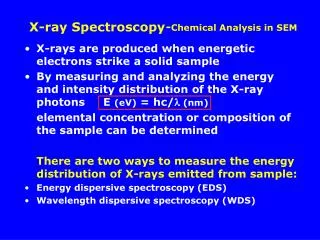
X-ray Absorption Spectroscopy • EXAFS (Extended X-ray Absorption Spectroscopy) oscillations in X-ray absorption Gives a Radial Structure. • Examine Fourier transform – peaks occur at inter-atomic distances (usually not interpreted directly). • Fit theoretical model to EXAFS spectra. • Modern ab initio codes (e.g. FEFF) are very accurate – little requirement for standards. Graham N. George
X-ray Absorption Spectroscopy – Strengths and Limitations • Examines all of a particular element in a sample. • Can examine any phase (solids, solutions etc.). • Accurate bond-lengths (better than 0.02 Å). • Approximate coordination numbers & atomic number (15%). • Oxidation state information from near-edge (often only relative). • Poor distance resolution ΔR≈π/2k – generally about 0.15 Å. • Little or no geometrical information. • Analysis not always reliable (especially with “black box” software). Graham N. George
X-ray Absorption near-edge spectra – sensitivity 2- 2- selenate elemental Se selenite Se-methionine Graham N. George
Density Functional Calculations • Many modern codes are simple to use and run. • Inexpensive computer systems (e.g. we use an 8 x 2.8GHz Xenon processor Linux cluster). • EXAFS analysis runs on the same computers. • The absolute accuracy of bond-lengths is poor – computed bond-lengths are up to about 0.1 Å too long (with the functionals we used). Density Functional theory calculations used the Dmol3 Materials Studio V2.2. The Becke exchange and Perdew correlation functionals were used to calculate both the potential during the SCF, and the energy. Double numerical basis sets included polarization functions for all atoms. Calculations were spin-unrestricted and all electron core potentials were used. Graham N. George
DMSO reductase 2H+, 2e- H2O Prototypical member of the DMSO reductase family of Mo enzymes Catalyses the two-electron reduction of dimethylsulfoxide to dimethylsulfide. Graham N. George
DMSO reductase active site structure Oxidized enzyme has a mono oxo active site with two pterin dithiolenes coordinated. Graham N. George
DMSO reductase Active Site Structure - Perspective • Previously there has been much debate about the structure of the active site. • Crystal structures have been published with chemically impossible arrangements of atoms at the active site (e.g. active site too crowded). • Essentially all DMSO reductase crystal structures published to date have had some sort of problem of this nature. • These problems have been attributed to multiple species co-crystallizing. Graham N. George
DMSO reductase – interaction with substrates and products S Ser147 Bound DMSO O S Mo DMSO reductase binds dimethylsulfide to form a pink-purple species. The exact nature of this novel species is very interesting as it is likely to be important in developing an understanding of catalytic mechanism. McAlpine, A. S.; McEwan, A. G.; Bailey, S. (1998) J. Mol. Biol. 275, 613-623. Graham N. George
Interaction of DMSO reductase with dimethyl sulfide • Open questions: • Is it an oxidized or a reduced species? Suggestions include: • A fully reduced MoIV site.1 • A partly reduced site MoV-O-·S(CH3)2.2 • An oxidized MoVI site.3 • Is the S-O bond longer than normal? • Crystallography indicates 1.7 Å, which compares with the value of 1.53 Å for DMSO bound to Mo in models, and 1.50 Å for free DMSO. Suggested that binding to enzyme weakens the S=O double bond. Resonance Raman disagrees with this… • McAlpine, A. S.; McEwan, A. G.; Bailey, S. (1998) J. Mol. Biol. 275, 613-623. • Bray et al. (2001) Biochemistry40, 9810-9820 • Bennett, B. et al. (unpublished) Graham N. George
EXAFS of (CH3)2S bound DMSO reductase Mo-S + Mo-O data fit Mo-O EXAFS indicates 4 Mo-S at 2.37 Å 1 Mo-O at 2.23 Å 1 Mo-O at 1.98 Å • No short Mo=O bond (in disagreement with crystallography). • Cannot see distant sulfur of bound DMSO. George et al. (1999) J. Am. Chem. Soc. 121, 1256-1266. Graham N. George
Interaction with alternative products • EXAFS from heavier atoms (larger atomic number) is much easier to observe. • Product analogues tested with heavier atoms in place of sulfur. Dimethylsulfide – ~5mM (CH3)2S forms 100% DMSO bound form. Dimethylselenide – ~60mM (CH3)2Se forms ~50% analogous species. Trimethylarsine – 1:1 (CH3)3As (stoichiometric) with enzyme. Trimethylphosphine – ~5mM (CH3)3P yellow species forms. Graham N. George
Mo K near-edge spectra oxidized (CH3)2S (CH3)2Se (CH3)3As • All near-edge spectra are subtly shifted to lower energy with respect to oxidized enzyme. Consistent with a relative reduction of the metal site (e.g. MoIVvs. MoVI oxidized). Graham N. George
Mo K-edge EXAFS Fourier Transforms oxidized (CH3)2S (CH3)2Se (CH3)3As Mo-S Mo=O mono-oxo tetrathiolate des-oxo tetrathiolate species No EXAFS observed for (CH3)2S sulfur Mixed with oxidized enzyme, Mo···Se observed Mo····As stoichiometric, Mo···As observed • (CH3)2S, (CH3)2Se and (CH3)3As appear to form structurally related species. Graham N. George
As K near-edge spectra DMSOR + (CH3)3As (CH3)3As (CH3)3AsO • Arsenic is oxidized to AsV in (CH3)3As bound enzyme Graham N. George
As K-edge EXAFS As-C data As As=O fit As····Mo fit data Mo-S Mo-O Mo····As Mo • EXAFS shows (CH3)3As located at Mo site. • Both As=O and As-C interactions are clearly resolved. Graham N. George
EXAFS of (CH3)3As-bound DMSO reductase 1.91 Å As 1.70 Å Ser147 3.44 Å O 2.23 Å 2.01 Å Mo S 2.37 Å • Arsenic is oxidized (AsV) • Molybdenum is reduced (MoIV) • As=O bond-length is within normal range – no particular distortion is present. Graham N. George
DFT of (CH3)3As-bound DMSO reductase • (CH3)3As remains bound but with longer than observed Mo-O=As distance. • DFT Mo-S 2.41, Mo-O(Ser) 1.95, Mo-O(AsMe3) 2.45, Mo-As 3.56 • EXAFS Mo-S 2.37, Mo-O(Ser) 2.01, Mo-O(AsMe3) 2.23, Mo-As 3.44 Graham N. George
DFT Calculation – (CH3)2S=O leaves active site… • DMSO dissociates – rather than DMS – tendency to go in reverse. • Active site pocket must be important in stabilization and direction. • Future calculations will use hybrid approach to include protein. Graham N. George
Calculations suggest that (CH3)2S and (CH3)3As might behave differently… • (CH3)2S should bind less tightly than (CH3)3As – biochemical data are consistent with this. • (CH3)2S-DMSOR gives a transient Mo(V) EPR with PES oxidation. With (CH3)3As-DMSOR the oxidation occurs too quickly to readily observe Mo(V). • (CH3)3As-DMSOR has a “normal” As=O bond, but does (CH3)2S? • Mo K near-edge spectrum of (CH3)3S-DMSOR is almost identical but very slightly higher in energy than (CH3)3As-DMSOR. • Next step – examine sulfur K-edge spectra of DMS-bound enzyme (very difficult to do – there’s lots of other sulfur there…). Graham N. George
(CH3)2S-DMSO reductase – Conclusions. • Mo site is best described as a reduced, formally Mo(IV), site. • (CH3)3As data suggest “normal” As=O and, by inference, S=O bond-lengths in product-bound enzyme species. • Calculations suggest that there may be some differences in the behavior of (CH3)2S and (CH3)3As. • Future work: • Use sulfur K-edge to understand the true nature of (CH3)2S binding. • Explore effects of protein by incorporation into calculations (hybrid approach). Graham N. George
The Future – High Resolution EXAFS • A major limitation of EXAFS is that it has poor distance resolution. • In principal this is simple to address – significantly increase the range of the data. • Goal – improve distance resolution to below 0.1Å – and resolve individual bonds to molybdenum. Graham N. George
Effect of k-range on EXAFS resolution Mo N N Mo Graham N. George
Sulfite Oxidase H2O 2H+, 2e- HSO3- HSO4- • Catalyzes the oxidation of sulfite to sulfate. • Sulfite Oxidase Crystal Structure • Initially, the enzyme was in the fully-oxidized MoVI form • Photoreduction (probably) during data acquisition reduced enzyme to MoIV via MoV. • Data likely arises from of a mixture of all three oxidation states. Graham N. George
Sulfite Oxidase reduced oxidized S O S O Mo S(Cys) Mo S(Cys) S O S O • Oxidized enzyme is a cis-dioxo MoVI site with one molybdopterin dithiolene ligand and one protein cysteine ligand. • Reduced enzyme is a mono-oxo MoIV site one Mo-OH or Mo-OH2ligand. Graham N. George
High resolution EXAFS of sulfite oxidase 2002 9-3, 55min. k=25 Å-1 k3~15,600 High-resolution EXAFS Scan 1996 7-3, 35min. k=14 Å-1 k3~2,700 Ordinary EXAFS Scan • Modern high-intensity beamlines and detector systems allow us to significantly extend the range of the data. • This allows data to be collected at higher resolution. • Technical issues : Problems with data acquisition (beamline stability). Problems using ab initio theory at very high k. Graham N. George
High resolution EXAFS of sulfite oxidase Mo=O Mo-S oxidized data fit reduced • Oxidized Enzyme – data extends to k=25.0 Å • Reduced Enzyme – data extends to k=21.5 Å Graham N. George
High-resolution EXAFS of Oxidized Sulfite Oxidase 1.72 2.42 2.42 1.72 2.42 • Individual Mo-S and Mo=O cannot be resolved with high-res. EXAFS. • DFT suggests that Mo=O bond-lengths should be equal. • Using the Mo-S Debye-Waller factor, we calculate that individual Mo-S distances must differ by 0.05Å or less. • DFT suggests that Mo-S bond-lengths differ by less than 0.05Å. Graham N. George
High-resolution EXAFS of Reduced Sulfite Oxidase 2.35 1.72 2.41 2.35 2.30 • One of three Mo-S bond-lengths is resolved. • Assignments based on DFT calculations. • The Mo-O bond at 2.30 Å is resolved from the Mo-S interactions. This bond-length indicates a bound water, rather than an -OH. Graham N. George
High resolution EXAFS spectroscopy • Experiments out to k=25 Å are quite feasible. • May need to go to even higher resolution (> k=30 Å). • This will require even higher intensity synchrotron beamlines (under development as we speak…), improved beamline stability, and possibly improved solid-state detector arrays. • At the molybdenum K-edge and using state-of-the art beamlines X-ray photo-damage is not a problem – but at some stage it will become so. Graham N. George
Graham George / Ingrid Pickering Group Graham N. George
Acknowledgements The National Institutes of Health GM57375 The Stanford Synchrotron Radiation Laboratory is a national user facility operated by Stanford University on behalf of the U.S. Department of Energy, Office of Basic Energy Sciences. The SSRL Structural Molecular Biology Program is supported by the Department of Energy, Office of Biological and Environmental Research, and by the National Institutes of Health, National Center for Research Resources, Biomedical Technology Program. Graham N. George
Future Directions… • Spectroscopy at low temperatures ! • As of Summer 2003, Graham George & Ingrid Pickering Canada Research Chairs in X-ray Absorption Spectroscopy and Molecular Environmental Science at University of Saskatchewan, home of the Canadian Light Source Graham N. George