Circulatory System Chapter 4
Circulatory System Chapter 4 . Veterinary Technology. Introduction. Vertebrates have a closed circulatory system, which generally confines blood within its walls. The closed systems coevolved with the respiratory and lymphatic systems. In fish, blood flows in one circuit.

Circulatory System Chapter 4
E N D
Presentation Transcript
Circulatory System Chapter 4 Veterinary Technology
Introduction • Vertebrates have a closed circulatory system, which generally confines blood within its walls. • The closed systems coevolved with the respiratory and lymphatic systems. • In fish, blood flows in one circuit. • In birds and mammals, it flows in two through a partitioned heart that works as two side-by-side pumps. • The double circuit supports the high levels of activity typical of most vertebrates that evolved on land.
Blood Components and Functions • When blood is spun in a centrifuge, the liquid and solids portions separate and the solids settle to the bottom. • This cellular portion makes up about 30% to 45% of total blood contents. • The remaining fluid is plasma.
what’s in digested food red blood cells white blood cells BLOOD oxygen waste (urea) platelets carbon dioxide hormones plasma
The Blood white blood cell red blood cell plasma platelets
Red Blood Cells – AKA Erythrocytes contain hemoglobin, a molecule specially designed to hold oxygen and carry it to cells that need it. a biconcave disc that is round and flat withoutanucleus can change shape to an amazing extent, without breaking, as it squeezes single file through the capillaries. An adult dog has about 6 to 8 million RBC’s per microliter!
Red Blood Cells – AKA Erythrocytes • Erythropoieses = the production on RBC’s by the bone marrow. • Erythropoietin = hormone produced in the kidney to stimulate RBC production. • When and why do animals need this?
White Blood Cells – AKA Leukocytes there are many different types and all contain a big nucleus. the two main ones are the lymphocytes and the macrophages. macrophages ‘eat’ and digest micro-organisms. some lymphocytes fight disease by making antibodies to destroy invaders by dissolving them. other lymphocytes make antitoxins to break down poisons.
Blood Components and Functions • Types of White Blood cells – page 48 • Types of Blood proteins – page 46
Platelets Platelets are also produced in the bone marrow, help with blood clotting. Platelets work with the protein Fibrinogen Platelets produce tiny fibrinogenfibersto form a net. This net traps other blood cells to form a blood clot. What is the process of blood clotting?
Plasma – about 94% water It also contains useful things like; • carbon dioxide • oxygen • nutrients • glucose • electrolytes • proteins – about 8% • minerals • vitamins • hormones • waste materials • like urea. A straw-coloredliquid that carries the cells and the platelets which help blood clot.
Directional Terminology • Helpful to know, when describing anatomy and describing blood flow.
Medial refers to being directed toward the middle or median plane of the body. • The median plane is best described as a line that would divide the body in half from head to tail. • 2. Lateral refers to being directed away from the median plane of the body
1. Cranial is directed toward the head or cranium. • It is used on the legs to describe structures above the metacarpal/ankle/fetlock joint. • Dorsal is used to describe structures below the aforementioned joint, along with palmar or plantar. • 2. Caudal is directed toward the rear of the body. • It is used for structures on limbs above the metacarpal/ankle/fetlock joint.
1. Dorsal refers to a body part that is directed toward the back of the body. • For instance, the hip joint is dorsal to the knee joint. • 2. Ventral refers to a body part that is directed toward the belly. • For example, the elbow joint is ventral to the shoulder joint
1. Proximal is directed toward the body from foot to head. • 2. Distal is directed away from the body from head to foot
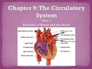
Blood Vessels and Blood Flow • In Mammals, The heart is a four-chambered, hollow muscle used to pump blood. • It provides two separate paths. • 1. Pulmonary Circuit: moves blood to lungs. Right Ventricle is the pump 2. Systemic Circuit: moves blood to the rest of the body. Left Ventricle is the pump
Blood Vessels and Flow • Page # 52 may be more helpful than my description. • Be sure and read this section! (pages 50 – 54)
Lungs Body cells Mammal’s circulatory system is a double circulatory system. This means it has two parts . the right side (Pulmonary) of the system deals with deoxygenated blood. the left side (Systemic) of the system deals with oxygenated blood.
blood from the lungs blood from the body The heart beat begins when the heart muscles relax and blood flows into the atrium. The relaxation phase is called the diastole phase How does the Heart work? STEP ONE: The Cardiac cycle
The atrium then contract and the valves open to allow blood into the ventricles. The contraction phase is called systole. How does the Heart work? STEP TWO: The Cardiac Cycle
The valves close to stop blood • flowing backwards. • The ventricles contract forcing • the blood to leave the heart. • At the same time, the atria are • relaxing and once again filling with • blood. How does the Heart work? STEP THREE: The Cardiac Cycle The cycle then repeats itself.
Mammalian Heart Structures • The heart is located low in the chest between the two lungs, and is contained within a fine membrane, called the pericardium. • The wall is mainly composed of cardiac muscle, the myocardium. • Smooth epithelium tissues lines both the inside and outside of the myocardium.
There are 3 types of blood vessels a. ARTERY b.VEIN c.CAPILLARY
The ARTERY Arteries carry blood away from the heart. Break down into smaller arterioles as they reach the lungs the elastic fibres allow the artery to stretch under pressure the thick muscle can contract to push the blood along. thick muscle and elastic fibres
The VEIN Veins carry blood towards from the heart. veins have valves which act to stop the blood from going in the wrong direction. thin muscle and elastic fibres body muscles surround the veins so that when they contract to move the body, they also squeeze the veins and push the blood along the vessel.
The CAPILLARY Capillaries link Arteries with Veins they exchange materials between the blood and other body cells (tissues) the wall of a capillary is only one cell thick The exchange of materials between the blood and the body can only occur through capillaries.
The CAPILLARY A collection of capillaries is known as a capillary bed. artery vein capillaries body cell
SUMMARY copy and complete the following; Arteries take blood ______ from the heart. The walls of an artery are made up of thick _________ walls and elastic fibres. Veins carry blood ________ the heart and also have valves. The _________ link arteries and veins, and have a one cell thick wall. Blood is made up of four main things ______, the liquid part of the blood; Red Blood Cells to carry ______; White Blood cells to protect the body from disease and _________ to help blood clot. away muscular towards capillaries plasma oxygen platelets
Electrocardiograms, Heart Sounds and Blood Pressure • The rate at which the heart beats is controlled by the nervous system. • Pacemaker cells (system): the cells that begin the heartbeat, and help control the rhythm. • AKA: Sinoatrial (SA) node • Found in the right atrium
Blood Pressure • The highest numbers is both the systole and diastole phases. • Measured in millimeters of mercury • Typical human is 120/80 mm Hg. • Systolic over diastolic pressure • Doppler machine • http://www.youtube .com/watch?v=no7Z8H8yCyQ
Blood Pressure • Ways to regulate BP: • BP is higher is arteries than veins • Page 59 • Kidneys produce a enzyme called renin when a low BP is detected. • BP is lower in the limbs (fight gravity) • Why do you faint when you lock your knees?
Electrocardiograms, Heart Sounds and Blood Pressure • Electrocardiograph – the instrument that picks up the electrical signal running through the body • Electrocardiogram – (ECG) the tracing made by the instrument. • Identifies problems associated with the contraction of the heart.
Electrocardiograms, Heart Sounds and Blood Pressure • sinus rhythm = Normal, consistent rate • Sinus trachycardia = faster than normal, but with a normal rhythm • Sinus bradycardia = slower that normal, but with a normal rhythm • Artial fibrillation = pacemaker not working, irregular • Asystole = flat line! • Cardiopulmonary resuscitation (CPR)
Electrocardiograms, Heart Sounds and Blood Pressure • Heart murmurs: defective valve or abnormal flow of blood. • Makes a swishing noise • In you notes: read page 56: Describe the typical heart noise and the action that happens for that noise to occur.
Clinical Practice • Blood analysis is the process of testing blood to determine its characteristics. • Samples are collected and tested to determine various attributes.
Science Starter • Read page 45 • What will happen to Sconic’s foot when the blood supply was stopped? • For a short period of time • For a long (several days) period of time?
Clinical Practice • Blood typing is a kind of analysis that is used in genetic studies. • Genetic markers in the blood can be used to determine the parents of offspring. • For example, the identity of the sire of a calf can be determined by blood typing a calf. • Newer DNA analysis procedures may be replacing some uses of blood typing.
Capillary Refill Time (CRT) • Check on gums, tissues under nails. • 1. Apply pressure for 5 seconds • 2. remove the pressure • In a healthy animal , the color should return to pink in just one second!
Clinical Practice: vocab! • Hypo- (value is below normal) • Hyper- (value is above normal) • -enima(used to describe levels in the blood stream • -penia (WBC count less than normal) • -cytosis (WBC count higher than normal) • -ia (other cells are elevated) • Erythro- (referring to RBC) • Auto- (referring to self)
Anemia • Low RBC count • Supplies less oxygen to the tissues • Signs: fatigue during exercise, weak, sluggish, inactive. • Not an actual disease – sign of other diseases • Causes: • 1. excessive blood loss • 2. shortened life of RBC • 3. decreased production of RBC • Parasites??? (hookworm) • Acetaminophen (Tylenol) • Autoimmune Disease