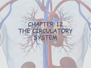
Chapter 9 Circulatory system
Chapter 9 Circulatory system. ---Closed tubular system ---Blood circulatory or cardiovascular S ---Lymph vascular S. The cardiovascular S heart artery vein capillary. 1. Capillaries. 1) LM : endothelium: basement membrane: pericyte: /flattened with processes /function:


Chapter 9 Circulatory system
E N D
Presentation Transcript
---Closed tubular system ---Blood circulatory or cardiovascular S ---Lymph vascular S
The cardiovascular S • heart • artery • vein • capillary
1. Capillaries 1) LM: • endothelium: • basement membrane: • pericyte: /flattened with processes /function: i. support ii. undifferentiated cell
endothelial cell: • processes –microvilli-like, finger-liked • vesicle /60-70nm, constitute about 25-35% of total volume /transendothelial channel function: transport large molecules and storage of membrane (for enlarge, enlongated, pore-formed and microvilli)
EM: classification ①Continuous capillary: ---Structural feature: • endothelial cell: -more vesicles -tight junction • basal lamina: complete ---Distribution: CT, MT, lung and CNS
② Fenestrated capillary ---Structural feature: • endothelial cell: -have fenestrae or pore (60-80nm in D, with 4-6 nm diaphragm) • basal lamina: complete ---Distribution: gastrointestinal tract, endocrine gland and renal glomerulus
③Sinusoid capillaries(enlarge capillary) ---Structural feature: • endothelium: gap, pore • basal lamina: incomplete or no (absent) ---Distribution: liver, spleen and bone marrow
2. Artery ---large A: aorta, pulmonary trunk ---medium-sized A: all named A, the diameter > 1mm (radial A, ulnar A) ---small A: 300um<D<1mm
Medium-sized A – muscular A ---Tunica intima • endothelium • subendothelial layer: CT -collagenous F -elastic F -smooth muscle • internal elastic membrane: wave-liked, pink-colored band- elastin
---Tunica media: thickest layer • smooth muscle: 10-40 layers of, circularly • elastic F: produced by SM • collagenous F: produced by SM ---Tunica adventitia: • LCT –with small BV-vasa vasorum, NF and LV • external elastic M
2) Large A: elastic A ---Structural features: a. Tunica intima is thick, and internal elastic M is not prominant b. Tunica media: consists of 40-70 layers of elastic membrane, CF and SM
SM elastic M Large A
3) Small A: ---large SA: • internal EM • 3-4 layers SM • no external M ---small SA: • no internal M • 1-2 layers SM
2. Veins ---three type ---correspond with A except for LV ---three layers
---structural features: a. larger diameter, thinner walls- collapsed b. no internal and external elastic membrane, so the boundaries between three tunica are not very clear c. contains more CT, less smooth M, SM are arranged in bundles d. vein valve: /infolding of tunica intima /semilunar-liked /prevent back flow of blood
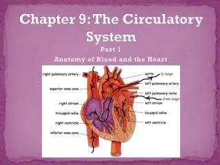
4. Heart ---atrium(right and left) ---ventricle (right and left)
E The wall of heart SE endocardium subendocardial layer myocardium epicardium
1) The wall of heart ---endocardium: • endothelium • subendothelial layer: more dense CT- contain fibroblast, CF, EF, SM • subendocardial layer: LCT, with BV, N and conducting S- Purkinje fiber
---myocardium: • cardiac M: a. atrial muscle: /atrial granules: 0.3-0.4um in D, contain atrial natriuretic polypeptide or cardionatrin b. ventricular M: thick, long, branches • LCT: rich in capillaries *atrioventricular fibrous annulus: DCT
---epicardium: visceral layer of pericardium- serous membrane: • mesothelium • LCT: more fat cell- subepicardial layer ---cardiac valve: • formed by infolding of endocardium: endothelium + DCT • prevent the back flow of blood
2) Conducting S ① components: ---sinoatriol node (SA node): located in epicardium of right atrium ---atrioventricular node (AV node) and bundles (AV bundles): located in subendocardial layer ---network of Purkinje fiber:
② three types of cells a. pacemaker cell ( P cell): • mainly distributed in SA and AV node • small, fusiform or polygonal in shaped • enclosed by DCT • less organelle: myofibril, plasmalemmal vesicles and more glycogen
b. transitional cell: • mainly distributed in periphery of SAN or AVN and AV bundle • The structure is between pacemaker cell and cardiac M • thinner and shorter than CM • more myofibril than P cell
c. Purkinje cell: • mainly constitute AV bundle and branches • shorter, boarder than CM, with 1-2 centrally located nuclei • rich in mitochondria, glycogen, less myofibril • well-developed intercalated disks
1. Components 1) Cells
① Lymphocyte: a. T-lymphocytes: • cytotoxic T cell: Tc C- kill the tumor cell, virus infective cell and foreign cell • helper T cell: Th C- promotes activity of BLC and Tc C • suppressor T cell: Ts C –regulate the function of BLC and TC b. B-lymphocytes: become into plasma cell c. NK cell: counteract virus infective cell and tumor cell
②Plasma cell ③antigen presenting cell: a. dendritic cell: • Blood DC • Langerhans cell • interstitial cell • veiled cell • interdigitating cell b.macrophage: Mononuclear phagocytic system
④other cells: • granulated cell • mast cell • blood platelet • blood-borne stem cell *Function: i. immunologic defence function ii. immune surveillance function iii. immune homeostasis
2) Lymphoid tissue ---reticular T: • reticular cell: stellate-shaped with processes to form network • reticular fiber ---lymphocytes, macrophage, plasma cell and mast cell
a. Diffuse LT: • no clear boundary • mainly consists of TLC • postcapillary venules: -high endothelial venules -opening for LC enter LT from blood
b. Lymphoid nodule: • spherical or ovoid • have clear boundaries • mainly composed of BLC • germinal center: stained pale * primary LN → secondary LN
3) Lymphoid organs • Central lymphoid organs: thymus, bone marrow • developed earlier • blood-borne stem cell comes from yolk sac • microenvironment → proliferation promoting • send LC to PLD and LT two weeks before borne
b. Peripheral lymphoid organs: lymph node, spleen and palatine tonsil • developed later • LC come from CLO • cell proliferation need antigen stimulating - antigen dependent • place for immune reactions