The Auditory and Vestibular Systems
450 likes | 694 Vues
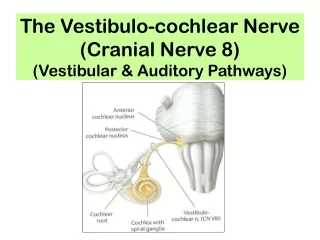
The Auditory and Vestibular Systems. I. Functional Anatomy of the 2 Systems - Overview Parallel ascending auditory pathways. Ascending vestibular system. II. Regional Anatomy Sensory organs 1. Auditory receptor cells and apparatus. 2. Vestibular sensory organs. B. Brainstem nuclei

The Auditory and Vestibular Systems
E N D
Presentation Transcript
I. Functional Anatomy of the 2 Systems - Overview • Parallel ascending auditory pathways. • Ascending vestibular system. II. Regional Anatomy • Sensory organs 1. Auditory receptor cells and apparatus. 2. Vestibular sensory organs. B. Brainstem nuclei 1. Vestibular and its projections. 2. Auditory and its projections. a. Lateral lemniscus b. Inferior colliculus c. Medial geniculate nucleus d. Primary auditory cortex. e. Wernike’s Area – a higher-order auditory cortex.
I. Functional Anatomy of the 2 Systems - Overview • Auditory system. • Tonotopic basis for frequency coding along the length of the cochlea. • Tympanic membrane transduced info into neural signals. (next slides)
Hearing • Sounds are waves of compressed air traveling through space - sound intensity wave height - pitch wave frequency
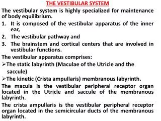
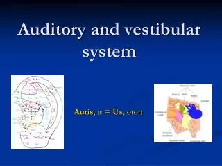
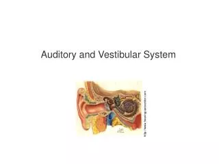
Cochlea Vestibular apparatus Organ of hearing (and equilibrium) – inner ear
1- The sound waves enter the external auditory canal and trigger vibrations of the tympanic membrane 2- The tympanic membrane induces a vibration of the ossicles 3- the last ossicle, the stapes, transmits amplified vibrations to the oval window 4- The vibrations induce waves in the perilymph of the various inner ear chambers 5- the round window absorbs excess energy and prevent wave reverberation 6- the fluid wave is transduced into an electrical signal by the auditory receptors, the organs of Corti located on the basilar membrane Sensory Organs: Hearing
The hair cells of the organ of Corti transduce fluid wave into an electrical signal The energy of the wave causes the basilar and vestibular membrane to move, thus displacing the cilia from the organ of Corti Receptors for sound: the organ of Corti
Movements of the cilia open or close potassium channels changes in the state of polarization of the hair cell Changes in potassium leakage due to cilia bending trigger changes in neurotransmitters exocytosis The neurotransmitters send an electrical signal to an afferent neuron of the cochlear nerve The louder the sound, the more the cilia bend, the more numerous are the APs produced Signal transduction
The location of the organs of Corti on the basilar membrane codes for pitch - Organs of Corti located near the oval window are more sensitive to high pitch sounds while the ones located toward the tip of the cochlea respond more readily to low pitch sound Coding for pitch
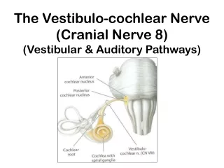
Neural pathway for sounds • Cochlear nerve nucleus in medulla oblongata thalamus auditory cortex in the temporal lobe • So, how do we perceive the direction from which a sound is coming from?
B. Brainstem nuclei and their Projections 2. Auditory nuclei: 3 major auditory relay nuclei of the brainstem: A. cochlear nuclei (same side from cn VIII) (medulla). B. superior olivary nuclear complex (integration from both sides) (pons). C. inferior colliculus (midbrain). [2 major divisions we noted earlier]. -superior olivary complex is important in sound localization (major input from AV cochlear nucleus).
Auditory System • Overview: Cochlear division of • Nerve VIII cochlear n. (same side) in rostral medulla. • 1. *Anteroventral cochlear n. • sup olivary n. (both • sides) lateral lemniscus • inferior colliculus. • *Important for horizontal location of sounds, as well as for other aspects of sound patterns, other than location. • 2. Dorsal posteriorventral • cochlear n. lat lemniscus • Inferior colliculus (opp. • side). • Note the decussations: Important for integration. • Clin: one can experience loss in only 1 ear only if lesion is peripheral.
B. Brainstem nuclei and their Projections 2. Auditory nuclei (cont’d): • Low-freq sounds are distinguished in space by interaural time difference. • High-freq sounds are distinguished by difference in intensity between the ears. • Different parts of the superior olivary n. (medial and lateral) are sensitive to these 2 types of differences. • Decussation is visible in trapezoid body. • Feedback pathway: some superior olivary neurons project back to the cochlea (both sides).
B. Brainstem nuclei and their Projections 2. Auditory nuclei (cont’d): • Olivocochlear bundle – regulates flow of auditory info to the brain (much like inhibitory dorsal horn n. inhibit somatic sensory info.). Lateral Lemniscus Most auditory path neurons course in lateral lemniscus inferior colliculus. Some synapse on nucleus of lateral lemniscus contralateral inferior colliculus. Another important site of decussation (Probst’s Commisure)
B. Brainstem nuclei and their Projections 2. Auditory nuclei (cont’d): Inferior Colliculus • Within the midbrain tectum. • The central nucleus within the inf colliculus receives the auditory info, which will proceed to the medial geniculate nucleus of the thalamus and the 1° auditory cortex. • Laminated – neurons in a single lamina are maximally sensitive to similar tonal frequencies. • Receives input from superior olivary n., n. of lateral lemniscus (both sides), and dorsal n. + pv cochlear (direct). • Projects to thalamus through the brachium of the inferior colliculus.
B. Brainstem nuclei and their Projections 2. Auditory nuclei (cont’d): Medial geniculate nucleus • Thalamic auditory relay nucleus. • The major part (ventral division) is tonotopically organized (receiving its input from the central n. of the inf coll, which is also tonotopically organized. • Therefore, the MGN is also laminated – layers maximally sensitive to similar frequencies. • Thalamocortical auditory projections are called auditory radiations 1° auditory cortex with 2 gyri within sulcus of temporal lobe: Heschle’s Gyri
B. Brainstem nuclei and their Projections 2. Auditory nuclei (cont’d): Medial geniculate nucleus • Columnar organization of neurons sensitive to tones of similar frequencies (isofrequency columns). • Also binaural columns (similar interaural intensity differences – for localization of high-frequency sounds). • Like other 1° sensory cortices, this has a prominent layer 4.
B. Brainstem nuclei and their Projections 2. Auditory nuclei (cont’d): Wernicke’s Area • A higher-order auditory cortex for the interpretation of language. (language on L side of brain; interpreting emotional content of language on R side of brain). • One projection of Wernicke’s area is to Broca’s motor speech area in the frontal lobe.
Auditory System • Overview: Cochlear division of • Nerve VIII cochlear n. (same side) in rostral medulla. • 1. *Anteroventral cochlear n. • sup olivary n. (both • sides) lateral lemniscus • inferior colliculus. • *Important for horizontal location of sounds, as well as for other aspects of sound patterns, other than location. • 2. Dorsal posteriorventral • cochlear n. lat lemniscus • Inferior colliculus (opp. • side). • Note the decussations: Important for integration. • Clin: one can experience loss in only 1 ear only if lesion is peripheral.
Sensory Organs: Vestibular: Ampulae of semicircular canals Maculae of utricle (linear acceleration) + saccule Endolymph – gel-like fluid flows over the hair cells with movement and deflects them – Ca carbonate crystals (otoliths)
Ability to detect head position and movement (or acceleration) Change of speed = linear acceleration (utricle and saccule) Turning = rotational acceleration (semi-circular canals) Equilibrium
Sensory cells have cilia extending into a gelatinous material topped by otoliths Saccule detects backward-frontward movement Utricle detects changes relative to gravity Utricle and saccule
The receptors in the ampulla are hair cells with cilia extruding into a gelatinous mass (cupula) When the head rotates, the cupula moves cilia pulled APs (vestibular nerve cerebellum …) Semi-circular canals
So why does a person become dizzy after he/she stops spinning?
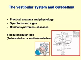
B. Brainstem nuclei and their Projections • Vestibular nuclei – on floor of 4th ventricle. 4 nuclei: inferior, medial, lateral, superior ascending projections to VPN of thalamus 1° vestibular cortex in parietal lobes (just behind the 1° somatic sensory cortex). Can project to nearby parietal areas for integration of info regarding head motion with info from somatic sensory receptors in trunk and limbs.
Vestibular System Overview: Head motion vestibular hair cell receptors 4 vestibular n. in rostral medulla and caudal pons: 2 Descending projections to sc and extraocular muscles (control movements) cerebellum. 2 Ascending projections VPN of thalamus 1° vestibular ctx in parietal lobe (for conscious awareness of orientation and motion).