Auditory and vestibular system
230 likes | 545 Vues
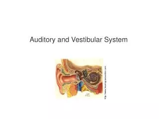
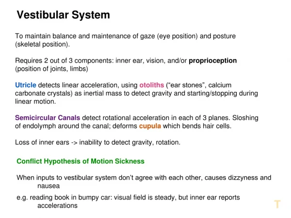
Auditory and vestibular system. Sensory organs on the inner ear. Eckert: Animal Physiology, W.H.Freeman and Co., N.Y.,2002, Fig. 7-27 a,b. inner ear: audition (exteroceptor) and vestibular apparatus (proprioceptor) bony and membranous labyrinths within the temporal bone ( os temporale )

Auditory and vestibular system
E N D
Presentation Transcript
Sensory organs on the inner ear Eckert: Animal Physiology, W.H.Freeman and Co., N.Y.,2002, Fig. 7-27 a,b. • inner ear: audition (exteroceptor) and vestibular apparatus (proprioceptor) • bony and membranous labyrinths within the temporal bone (os temporale) • both sensory organs contain hair cells: secondary receptor cells • these sensory systems are related evolutionary with the lateral-line organ (fish, amphibians) – acousticolateral organ • structure of hair cells is similar everywhere • hair cells are flanked by supporting cells – tight junctions: perilymph and endolymph separated • perilymph: extracellular space, high Na+, low K+ • endolymph: transcellular fluid, high K+, low Na+ • endolymph is positive compared to perilymph – 150 mV compared to the intracellular space • at the tip of the stereocilia mechanosensitive K+ channels, 10-15% open - 90 Hz firing rate on primary axons – changes depend on direction of bending 2/14
Structure of hair cells 3/14 stereocilium K+ tip-link kinocilium basal connection supporting cell hair cell supporting cell actin transmitter Na+ K+ ATP Ca+ K+ Na+ endolymphhigh K+ + 80 mV - 70 mV perilymphhigh Na+ + 0 mV
Cilia on hair cells 4/14 Eckert: Animal Physiology, W.H.Freeman and Co., N.Y.,2002, Fig. 7-24
Vestibular system I. 5/14 Eckert: Animal Physiology, W.H.Freeman and Co., N.Y.,2002, Fig. 7-27 c,d. Eckert: Animal Physiology, W.H.Freeman and Co., N.Y.,2002, Fig. 7-25 • vestibular apparatus consists of two parts: • 3 semicircular canals – detect angular acceleration • otolith organs (utriculus, sacculus) – detect position of the head, linear acceleration • semicircular canals are approximately perpendicular to each other • horizontal one is tilted downwards by 25°, the two vertical ones have an angle of 41° and 56°, respectively with the sagittal plane • sensory epithelium with the hair cells is called crista ampullaris, it is located at one end of the canal in the ampulla • hair cells are covered by the cupula that almost blocks the flow of the endolymph • lateral-line organ works similarly • kinocilia are uniformly oriented in the crista • semicircular canals are in pairs within the same plane - complementary pairs – when one is excited, the other is inhibited
Vestibular system II. 6/14 Eckert: Animal Physiology, W.H.Freeman and Co., N.Y.,2002, Fig. 7-27 c,d. • hair cells are located in maculas within the utriculus and sacculus • macula is horizontal in the utriculus, while it is vertical in the sacculus • hair cells are covered by otolith membrane containing otoliths (calcium carbonate) • kinocilia in maculas (maculae) are oriented with respect to an undulating line, the striola • any position of the head causes a specific firing pattern in the primary afferents • linear acceleration (lift) is also detected because of the inertia of the otoliths • primary sensory neurons are located in the ganglia vestibulare • central axons run to the four vestibular nuclei in the brainstem
Vestibular centers 7/14 • Deiters’ nucleus (nucl. vestibularis lateralis) • strong, tonic excitatory effect on spinal motor neurons – tractus vestibulospinalis lateralis • crucial for the maintenance of straight posture • opposite effects: cerebellar inhibition on Deiters’ nucleus, cortical (in tetrapods rubral) inhibition on spinal motor neurons • decerebration rigidity • nucl. vestibularis medialis • brief, short lasting input from semicircular canals – control of neck muscles • nucl. vestibularis superior • similar input – control of eye movements • nucl. vestibularis inferior • less known, it integrates inputs with cerebellar information and provides ascending pathways
Audition 8/14 • ear is one of the most important telereceptive organ – vision is limited by darkness, olfaction depends on the direction of the wind • audition is functioning even during sleep – weak sounds made by the baby awake the mother • audition is very important for communication too • sounds are longitudinal waves • human ear is sensitive to sounds between 20 Hz and 20 kHz, some animals hear ultrasounds (rat after coitus); infrasound is annoying • intensity in given as the logarithm of the ratio to a reference value (20 μPa – threshold at 2 kHz) because the range of intensities is wide • in practice tenth of Bel, decibel (dB) is used, thus the logarithm should be multiplied by 10 • if voltage or current is used, then the second powerof these values should be entered in the equation – that’s why we use 20 and not 10 in the equation
The organ of audition 9/14 Eckert: Animal Physiology, W.H.Freeman and Co., N.Y.,2002, Fig. 7-29. • human ear consists of 3 parts: external, middle and inner ear • external ear: • pinna (can be moved in some animals) • auditory canal • tympanic membrane – closing external ear • middle ear: • ossicles (malleus, incus, stapes) – 22-fold increase in pressure because the different surface size and the lever system • Eustachian tube to the pharynx – equalization of pressure in the external and middle ears (flying, yawning, candy) • inner ear: • bony and membranous labyrinth with three canals • scala media (ductus cochlearis) surrounded by membrana basilaris (with the organ of Corti) and Reissner’s membrane • above: scala vestibuli, below: scala tympani
Mechanism of audition I. 10/14 Eckert: Animal Physiology, W.H.Freeman and Co., N.Y.,2002, Fig. 7-30 a,c. • ossicles forward sound stimuli to the perilymph (scala vestibuli) through fenestra ovale • cochlea has 2,5 turns – sound waves reach the top of the cochlea, then return through scala tympani to fenestra rotunda • total length of the cochlea is about 32-33 mm • sound waves also reach inner ear by bone conduction – it is less important, except when listening to our own sound record, or when middle ear is damaged and hearing aid is needed • the site of maximal oscillation of the membrana basilaris depends on frequency – tonotopy • movement of membrana basilaris stimulates hair cells • membrana basilaris is narrow and tight at the base (100μ), and wide and loose at the top (500μ) • Helmholtz suggested, György Békésy proved that high pitch tones are detected at the base, low pitch tones at the top of the cochlea
Mechanism of audition II. 11/14 • function of inner and outer hair cells is different • inner hair cells – sensation, outer hair cells - setting sensitivity • activation induces shortening of outer hair cells due to activation of their cytoskeleton – amplitude of maximal oscillation increases • threshold of inner hair cells is higher, it reaches normal detection limit only due to amplification • noise-induced hearing loss: (Walkman), and certain medicines (streptomycin) destroy outer hair cells • sterocilia are also shorter and tighter at the base of the cochlea – contributes to tonotopy • outer hair cells reach tectorial membrane, inner ones not – turbulently flowing perilymph stimulates them
Mechanism of audition III. 12/14 • mechanosensitive channels of stereocilia respond with time resolution in the μs range – K+ enters, because of the 150 mV potential difference • depolarization causes Ca++ influx – shape change in outer hair cells, increased glutamate (?) release in inner hair cells • primary sensory neurons are located in the ganglion spirale – 1 hair cell 10 afferents, 1 afferent – 1 hair cell • there are approx. 3500-3500 hair cells, i.e. 60-70000 afferents on the two sides together • afferents cannot follow 20 kHz frequency, spikes appear phase-locked • intensity is coded partly in frequency, partly in the number of cells activated (population code) depending on recruitment of neighboring and high-threshold hair cells • sensitivity is set by the lateral (inner hair cells and primary dendrites) and medial (outer hair cells) olivocochlear bundle
Central auditory pathway I. 13/14 • onset and termination of sounds, location of the source, and stimulus pattern should be analyzed by the central apparatus • anatomy is known, physiology is less well • general characteristics: parallel ascending fibers, two-directional connections, tonotopy • first relay stations are the cochlear nuclei (anteroventral, posteroventral and dorsal) – strictly ipsilateral • deafness restricted to one side is either because of damage to this nuclei or disruption at the periphery • primary afferents related to one hair cell (approx. 10 axons) terminate in one layer • fibers originating in these nuclei, join ipsi- and contralateral lemniscus lateralis and go to inferior colliculus, or reach superior olivary nucleus – second relay station
Central auditory pathway II. 14/14 • oliva receives bilateral projection, it localizes sound source – based on differences in phase [below 2 kHz], or in intensity [above 2 kHz] • 1° difference in direction can be detected • fibers from oliva join lemniscus lateralis and run to the third relay station, inferior colliculus • inferior colliculus is also important in detection of sound direction – e.g. owls • output to non-auditory areas as well • fourth station is corpus geniculatum mediale(medial geniculate body) – tonotopic representation, input from other sensory areas as well • the end station is the primary auditory cortex, Br. 41-42 in the temporal lobe – several areas arranged in a tonotopic way
Structure of the inner ear Eckert: Animal Physiology, W.H.Freeman and Co., N.Y.,2002, Fig. 7-27 a,b.
Crista ampullaris and macula Eckert: Animal Physiology, W.H.Freeman and Co., N.Y.,2002, Fig. 7-27 c,d.
The lateral-line system Eckert: Animal Physiology, W.H.Freeman and Co., N.Y.,2002, Fig. 7-25
Structure of the labirynth Eckert: Animal Physiology, W.H.Freeman and Co., N.Y.,2002, Fig. 7-29.
Organ of Corti Eckert: Animal Physiology, W.H.Freeman and Co., N.Y.,2002, Fig. 7-30 a,c.