Download
1 / 34
440 likes | 790 Vues
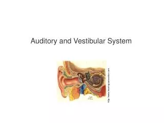
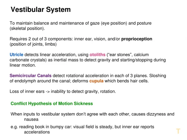
Vestibular System. Dr. G.R. Leichnetz. What does the vestibular system do ? It informs the brain of movement of the head and body: angular movement - semicircular canals linear movement - otolith organs (saccule & utricle)

E N D
Vestibular System Dr. G.R. Leichnetz
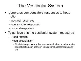
What does the vestibular system do? • It informs the brain of movement of the head and body: angular movement- semicircular canals linear movement- otolith organs (saccule & utricle) • When the head and/or body moves, the vestibular system: • Adjusts eye movements, compensating for head movement to maintain fixation, stabilize visual image (medial longitudinal fasciculus, ascending MLF, to extraocular motor nuclei). • Adjusts head position for “gaze” and balance (medial vestibulospinal tract, descending MLF, to cervical cord) • Adjusts posture for balance, equilibrium (lateral vestibulospinal tract) There is also vestibular input to the cerebellum, because the cerebellum coordinates the size and velocity of movements appropriate for the above circumstances. (vestibulocerebellar & cerebellovestibular fibers)
What is the Difference Between Proprioception and Vestibular Sense? Proprioception- position sensitivity- thru somatosensory receptors; position of the limbs, body in space Muscle spindles- stretch Pacinian corpuscles- pressure Meissner’s corpuscles- touch Vestibular Sense- movement sensitivity- thru vestibular receptors; change of head position, sense of balance, equilibrium Angular movement- semicircular canals Linear movement- otolith organs (saccule & utricle)
The semicircular canals are part of the membranous labyrinth located within the petrous portion of the temporal bone, situated in the three planes of orientation (anterior, posterior, horizontal canals).
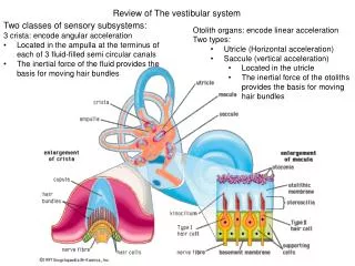
Vestibular Receptors: The vestibular end organs sense changes in the position and movement of the head in the three dimensions of space. Semicircular canals- crista ampullaris in ampullae angular movementOtolith organs- maculae of the saccule and utricle gravitation, linear movement Semicircular canals Otolith organs Netter Cochlea
Vestibular Receptors Semicircular canals: Crista ampullaris containing hair cells with kinocilium, stereocilia, covered by cupula (gelatinous cap) Otolith organs: Saccule & Utricle The maculae containing hair cells covered with gelatinous mass with otoconia Crista ampullaris of the ampullae of the semicircular canals Maculae of the saccule and utricle Netter
Crista Ampullaris, Semicircular Canals- detects angular movements Ampulla Hair cells Planum Semilunatum- produces endolymph
Maculae in Saccule &Utricle Hair cells, macula of utricle, cat Otoconia, macula utricle, cat Otoconia Hair Cells
When the stereocilia are displaced toward the kinocilium the hair cell is depolarized (excitation), and when displaced away from the kinocilium the hair cell is hyperpolarized (inhibition). Primary vestibular afferents end on hair cells
Crista Ampullaris of the Semi-Circular Canals Ampulla At Rest Ampulla During Movement Kandel, et al., The Principles of Neural Science
Primary vestibular fibers have their bipolar cell bodies of origin in the vestibular (Scarpa’s) ganglion and travel in the vestibular division of C.N. VIII to terminate in all subdivisions of the vestibular complex, and the flocculonodular lobe of the cerebellum. Flocculonodular lobe Vestibular receptors Vestibular complex Vestibular ganglion
The vestibulocochlear nerve (C.N. VIII) emerges with the facial nerve (VII) from the cerebellopontine angle next to the flocculus (hemispheric portion of the flocculonodular lobe). Facial nerve (VII) Cerebellopontine angle Vestibulocochlear nerve (VIII) Flocculus (flocculonodular lobe)
Vestibular signals are transmitted via the vestibular division of the vestibulocochlear nerve (C.N. VIII) to the vestibular complex. Lateral Recess of 4th ventricle Vestibular Complex ICP Vestibular Complex Flocculus ICP Vestibulocochlear Nerve (VIII)
The vestibular complex contains four subnuclei: superior, medial, lateral, and inferior vestibular nuclei MED INF DCN Inferior cerbellar peduncle Vestibulocochlear nerve (VIII)
The primary vestibular neurons have their bipolar cell bodies in the vestibular ganglion. Their peripheral processes end on the hair cells in the crista ampullaris or maculae of saccule & utricle. Their central processes travel in the vestibular division of the vestibulo-cochlear nerve (C.N. VIII) and terminate in all subnuclei of the vestibular complex. Some primary afferents go to the flocculonodular lobe of the cerebellum. Vestibular complex Vestibular Ganglion Netter
Second-order vestibular fibers originate from the vestibular complex and ascend in the medial longitudinal fasciculus (MLF) to the extraocular motor nuclei (III, IV, and VI) to coordinate the vestibulo-ocular reflex. The VOR produces eye movements to compensate for head movements, keeping the visual image on the fovea of the retina (maintaining fixation). III IV Medial longitudinal fasciculus VI Vestibular complex
Second-order vestibular fibers originate primarily from the medial and superior vestibular nuclei to ascend thru the medial longitudinal fasciculus to the extraocular motor nuclei. These fibers coordinate the vestibulo-ocular reflex. Canal-specific projections target oculomotor cell groups to orchestrate appropriate compensatory eye movements. From Spencer in: Conn, Neuroscience in Medicine
Vestibulospinal System The medial vestibulospinal tract (descending MLF) originates primarily in the medial vestibular nucleus and descends to the cervical spinal cord (vestibulo-colic reflex). The lateral vestibulospinal tract originates from the lateral vestibular nucleus and descends ipsilaterally in the anterior funiculus through the entire length of the spinal cord to terminate in the medial part of the ventral horn (vestibular influence on posture and equilibrium). Medial vestibulospinal tract (MLF) Lateral vestibulospinal tract
Medial vestibulospinal tract (MLF) to cervical cord to terminate on neck muscle motoneurons (vestibulocolic reflex). Adjustments in head position. Lateral Vestbulospinal tract is excitatory to motor neurons in the medial part of the ventral horn of the spinal cord, innervating axial and proximal limb musculature which affect posture and equilibrium. Medial vestibulospinal tract (MLF) ends in the cervical spinal cord Lateral vestibulospinal tract extends to entire length of the spinal cord
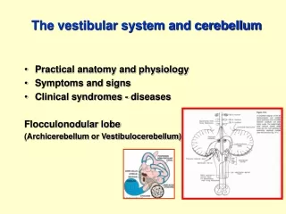
VESTIBULAR CONNECTIONS WITH THE VESTIBULAR PARTS OF THE CEREBELLUM: FLOCCULONODULAR LOBE AND FASTIGIAL NUCLEUS
The cerebellum receives proprioceptive and vestibular input and uses this sensory information to coordinate the appropriate size and velocity of movements. For example, in the VOR there must be a compensatory eye movement that precisely matches the head movement, but in the opposite direction. What parts of the cerebellum are associated with vestibular function?
The flocculonodular lobe (flocculus + nodule) is the vestibular part of the cerebellum, which is phylogenetically the oldest part of the cerebellum (archicerebellum). Flocculonodular lobe
The flocculus is the hemispheric part of the flocculonodular lobe of the cerebellum. Vestibulocerebellum The nodule is the vermal part of the flocculonodular lobe.
The vestibular portion of the cerebellum (flocculonodular lobe) receives some direct primary vestibular afferents. Flocculonodular lobe Vestibular complex Vestibular ganglion
Primary and secondary vestibulocerebellar fibers enter the cerebellum through the inferior cerebellar peduncle (juxtarestiform body) to terminate primarily in the flocculonodular lobe, the vestibular part of the cerebellum. Flocculonodular lobe Primary and secondary vestibulocerebellar afferents Vestibular nerve & ganglion
CEREBELLAR CORTEX Vestibulocerebellar fibers enter the cerebellum thru the inferior cerebellar peduncle (JRB) and end on granule cells in the cerebellar cortex (flocculonodular lobe), which project in turn to Purkinje cells. Purkinje cells of the cerebellar cortex project their axons to the fastigial nucleus, the vestibular-related deep cerebellar nucleus. Purkinje cells of the flocculonodular cortex project to the fastigial nuclei Primary vestibular afferents terminate on granule cells of the F-N cortex, which project in turn to Purkinje cells Fastigial nucleus
The fastigial nuclei are the medialmost of the deep cerebellar nuclei located in the subcortical white matter of the cerebellum in the roof of the fourth ventricle. These nuclei have reciprocal connections with the vestibular complex through the inferior cerebellar peduncle (juxtarestiform body). Fastigial nuclei
The fastigial nucleus, the medialmost of the deep cerebellar nuclei, has reciprocal connections with the vestibular complex thru the juxtarestiform body (part of inferior cerebellar peduncle). G F E D VC ICP Juxtarestiform body- part of the ICP that carries vestibulocerebellar and cerebellovestibular fibers F Dentate
While most primary vestibular fibers go to the vestibular complex, some go directly to the flocculonodular lobe of the cerebellum (vestibulo-cerebellum), which projects to the fastigial nucleus. The fastigial nucleus also has reciprocal connections with the vestibular complex. Fastigial nuclei Vestibulocerebellar and cerebellovestibular fibers traverse the juxtarestiform body
Feedback Projections from the Flocculonodular Lobe and Fastigial Nucleus to the Vestibular Complex Cerebellovestibular fibers traverse the juxtarestiform body.
What Clinical Signs Are Observed With Lesions of the Vestibular System? (eg. vestibular division of C.N. VIII, vestibular complex, MLF, flocculonodular lobe of cerebellum) 1. Vertigo- disruption of vestibular interaction with the visual system, resulting in a feeling that the room is spinning; dizziness. 2. Nystagmus- pathological nystagmus; rhythmic involuntary oscillation of the eyes; the eyes move slowly in one direction, and then jerk quickly back to the opposite side. 3. Loss of Balance, Equilibrium
Typically a benign tumor (proliferation of Schwann cells) that grows on the vestibular division of the vestibulocochlear nerve (C.N. VIII) in the cerebellopontine angle. A vestibular schwannoma can compress the vestibulocochlear nerve (C.N. VIII) with partial or complete deafness, tinnitis, vertigo, nystagmus, or facial nerve (C.N. VII) with weakness in ipsilateral face.