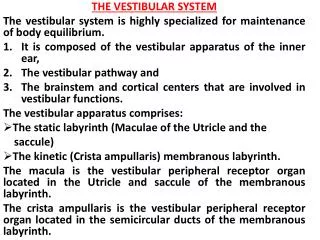
Vestibular system
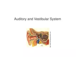
Part of the membranous labyrinth of the inner ear Involved in a form of proprioception The vestibular apparatus detects head movements and the position of the head in space - requires two sets of sensory epithelia to transduce angular and linear acceleration of the head


Vestibular system
E N D
Presentation Transcript
Part of the membranous labyrinth of the inner ear Involved in a form of proprioception The vestibular apparatus detects head movements and the position of the head in space - requires two sets of sensory epithelia to transduce angular and linear acceleration of the head - together they from five receptor organs (3 semicircular canals; as well as utricle and saccule) Receptive organs are ensheathed by connective tissue Vestibular system Subconscious motor
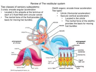
3 semicircular canals (anterior, posterior and horizontal) Respond to angular acceleration (yes, no, tipping of head) Utricle & Saccule Respond to linear acceleration & gravity Vestibular portions of inner ear: Membrane-lined fluid filled cavities in temporal bone (contains endolymph. connected with cochelar duct through ductus reuniens) Vestibular system Subconscious motor
Vestibular Components • A and P canals are oriented in vertical planes perpendicular to each other • H canal is oriented horizontally • sense events in 3 dimensions of space • Utricle & Saccule respond to linear acceleration & gravity • vestibular component of 8th cranial nerve; 20,000 myelinated axons Subconscious motor
Receptors = Hair Cells • - Depolarize when stereocilia are bent towards kinocilium (non-motile cilium; results in functional polarity of hair bundle) • Endolymph is high in K+; girdle of tight junctions separates endolymph from perilymph (like extracellular fluid; high in Na+) • afferents fire both tonically and phasically – firing can persist or adapt – resulting in mechanisms to signal sustained stimulation (acceleration from gravity) and abrupt changes in acceleration Subconscious motor
Respond to angular acceleration 3 on each side Filled with fluid Perpendicular to each other Pairs of canals in same plane Semicircular canals Subconscious motor
Mechanism of stimulation: Hair cells (7,000) located in ampulla - Gelatinous Cupula covers stereocilia (each “canal” is a closed tube of ~8 mm in diameter filled with endolymph) During rotation of head in the plane of a canal: Fluid moves around canal (acceleration detected by inertia) Fluid flow interrupted by cupula (Tilts the cupula; Stereocilia bent) Afferents excited on one side & inhibited on the other Semicircular canals Subconscious motor
Mechanism of stimulation: Fluid presses against one side of cupula Cupula bows, displacing the haircells Semicircular canals • All hairbundels share common orientation • Angular acceleration in preferred direction (towards kinocilium) depolarizes haircells and stimulates afferents, acceleration in opposite direction hyperpolarizes receptors • three canals are almost precisely perpendicular to one another • representing 3 mutually orthogonal axes Subconscious motor
Ovoidal sac of membranous laby-rinth about 3mm long - Utricle: 30,000 HC, saccule: 20,000 HC Respond to linear acceleration & gravity One of each on each side Utricle - macular surface horizontal Saccule - macular surface vertical Proportional activity in 2 channels for info on acceleration along all axes Mechanism of stimulation: hair cells in macular surface Stereocilia covered by gelatinous matrix Otoliths embedded in gelatin Otoliths more dense than water (fine, dense particles, “ear dust”) Mass lags behind movement of head Utricle and Saccule • gelatinous layer shifts with respect to underlying epithelium • deflects haircell bundles • elicits electrical response Subconscious motor
Utrical signals horizontal forces (utricle has variations in axes in populations of hair cells; tilt in any direction will depolarize some cells and hyperpolarize others) Saccule signals vertical forces Utricle and Saccule • - Linear acceleration or gravity forces otoliths to move gelatin and bend stereocilia Subconscious motor
Otoliths Utricle and Saccule Subconscious motor
Vestibulo-cochlear Nerve • nerve along which the sensory cells (hair cells) of the inner ear transmit information • consists of the cochlear nerve (hearing), and the vestibular nerve (balance) • emerges from the medulla oblongata and enters the inner skull via the internal auditory meatus in the temporal bone, along with the facial nerve. Subconscious motor
Vestibular information is used in 3 ways Control eye muscles so that in spite of changes in head position, the eyes can remain fixed on same point Reflex mechanisms for maintaining upright posture Conscious awareness of the potion and acceleration of body, perception of space surrounding the body and memory of spatial information Pathways Information is relayed from vestibular apparatus to nuclei in brainstem via vestibular branch of cranial nerve VIII Transmitted through multineuronal pathway through the thalamus to vestibular centers in parietal lobe and cerebellum descending projections sent to spinal chord to affect postural reflexes vestibular information integrated with info from joints, tendons and skin Vestibular Information and pathways Subconscious motor
2 vestibulospinal tracts (medial and lateral) Medial: Provides basic postural control receives much input from semicircular canals Causes movement of head and shoulders to coordinate head and eye movements (ends at cervical cord) Descend in the ipsilateral column of spinal cord; terminate in ventro-medial spinal gray matter; innervate axial and proximal muscles Vestibulo-Spinal Tracts; vestibulo-spinal reflexes Subconscious motor
2 vestibulospinal tracts Lateral: Concerned with goal-directed limb movement such as reaching and manipulating receives much input from utricle and saccule Changes muscle tone in response to gravity Descending pathway descend to dorsal part of lateral column of spinal cord Vestibulo-Spinal Tracts; vestibulo-spinal reflexes Subconscious motor
Other vestibular pathways ascend to oculomotor nuclei: CN III (oculomotor nerve; controls most of the eye's movements, constriction of the pupil, and maintains an open eyelid), CN IV (trochlear nerve; innervates a single muscle: the superior oblique muscle of the eye), CN VI (abducens nerve; controls the movement of a single muscle, the lateral rectus muscle of the eye) Cause eye movement in response to head rotation: Nystagmus Vestibulo-Spinal Tract: vestibulo-ocular reflexes Subconscious motor
Vestibular connections (postural control [medial] and limb movement [lateral]) to the cerebellum Thalamic information relayed to cortex - allow for conscious perception of head position and movement Projections from Vestibular Nuclei; vestibulo-spinal reflexes Subconscious motor
Afferent fibers relay through 4 vestibular nuclei (superior, lateral, medial and inferior) 2 vestibulospinal tracts Lateral: receives much input from utricle and saccule Changes muscle tone in response to gravity Medial: receives much input from semicircular canals Causes movement of head and shoulders to coordinate head and eye movements Strong input to cerebellum Central vestibular connections Subconscious motor
Together, vestibular reflexes stabilize eyes and body when head moves Vestibulospinal reflexes enable skeletomotor system to compensate for head movement Vestibuloocular reflexes keep eyes still when head moves Central vestibular connections Subconscious motor
Nystagmus; vestibulo-ocular reflexes Stabilize eyes when head moves Subconscious motor
Nystagmus; vestibulo-ocular reflexes • Stabilize eyes when head moves • you can read a book while shaking your head if the book is still (visual processing slower than vestibular processing for image stabilization) • vestibular apparatus signals how fast head is moving, ocular motorsystem uses info to stabilize eyes (visual image motionless on retina) • slow eye movement in opposite direction of head movement (driven by vestibular system; otholith reflex) • nystagmus to reset to center of gaze (driven by brain stem circuits) Subconscious motor
Subject seated on stool and rotated to left Initial response (hard to visualize) Slow tracking eye movements to right Fast eye movements back to left Nystagmus: alternate slow (otolith reflex) and fast eye movement (brain stem) Semicircular canals habituate, eyes begin to move in space Response to stopping turning (post-rotatory) Head stops but fluid continues moving left Eyes track slowly left, quick movement to right Nystagmus normal for head rotation and repetitive moving object (optokinetic) Nystagmus without movement = sign of lesion Vestibulo-occular control Post-rotatory nystagmus Subconscious motor
Vestibulo-occular control • Coffee cup example: • gently twist your coffee; watch a bubble at fluid boundary • at beginning, coffee tends to maintain its original orientation and thus counter rotates the cup • at conclusion of turning, when cup decelerates, coffee moves in opposite direction (post rotatory nystagmus) Post-rotatory nystagmus Subconscious motor