Vestibular System
Vestibular System. Lecture Outline. Signal Transduction Linear Acceleration and Angular Acceleration Vestibular Pathways Vestibulo-Ocular Reflex (VOR) Clinical Correlations. 2. Anatomy Summary: The Vestibular Apparatus .

Vestibular System
E N D
Presentation Transcript
Lecture Outline • Signal Transduction • Linear Acceleration and Angular Acceleration • Vestibular Pathways • Vestibulo-Ocular Reflex (VOR) • Clinical Correlations 2
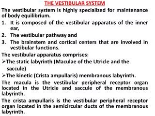
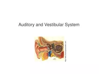
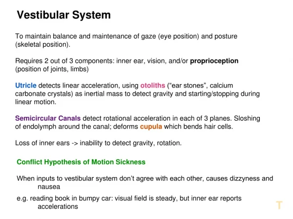
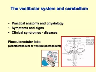
Anatomy Summary: The Vestibular Apparatus Vestibular apparatus provides information about movement and position of body in space- EQUILIBRIUM.
Hair Cells are the Transducers of the Inner Ear • The apex of a hair cell is the • signal transduction site • which contains cilia (mostly • stereocilia). • This area is surrounded by endolymph, • which has a higher concentration of potassium ions, compared to the usual extracellular fluid (or the • perilymph of the inner ear), is essential for the signal transduction process. • At the base of a hair cell • we find a synaptic terminal, with vesicles • containing excitatory transmitter. Synaptic • transmission between hair cells stimulates the afferent fibers of the vestibular and cochlear portions of the vestibulocochlear nerve (CN VIII).
Opening of mechanically gated Potassium Channels causes Depolarization of the Hair Cells • Mechanical force produced • by a “tip link” between neighboring stereocilia • directly opens the cation channels during deflection of the cilia towards the • tallest cilium(kinocilium). The • following inward current of potassium ions depolarizes the hair cells. Depolarization of Hair Cells increases Intracellular Calcium and induces Transmitter Release
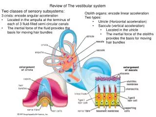
Two Types of Receptor Organs of the Vestibular System: Otolith Organs and Semicircular Canals Otolith Organs The saccule and the utricle are endolymphfilled pockets that contain in their walls a patch of sensory hair cells(maculae). Their cilia support, at their tips, calcium carbonate crystals glued together with a jelly like material (otolithicmembrane). When the cilia of the sensory hair cells are tilted, either by gravity forces, or by linear acceleration, the cilia bend, which leads to depolarization or hyperpolarization of the hair cells. Semicircular Canals -(horizontal, anterior vertical and posterior vertical), endolymph filled pipes which contain a patch of sensory hair cells (crista), with their cilia inserted into a jelly like structure (cupula). When the endolymph moves relative to the walls of the canals, induced by angular acceleration (rotation) of the head around one of the axes of the semicircular canals, the cupula with the inserted cilia bends, which leads to depolarization or hyperpolarization of the hair cells.
Rotational Forces in the Cristae The semicircular canals sense rotational acceleration The inertia of the endolymph inside the semicircular canal delays its participation in the rotation. It causes it to stay in its original position for a while, rather than following the rotation of its container (the semicircular canal) immediately. The endolymph flow, which occurs relative to the semicircular canal, produces pressure onto the cupula and causes it to bend. This also bends the cilia of the hair cells embedded in the cupula, and the hair cells, depending on their orientation, get either excited or inhibited.
Otolith Organs The otolith organs sense linear acceleration and head position
The Essence of Vestibular Pathways Most of the vestibular sensory information forms the afferent limb of vestibular reflexes (vestibulo-ocular and vestibulo-spinal reflexes). Vestibular reflexes stabilize the eyes and the body when the head moves. Some of the vestibular projections reach autonomic control centers in the hypothalamus. They are thought to be responsible for some of the symptoms of vestibular dysfunction like nausea, sweating, and increased heart rate, which can be perceived consciously
Types of Eye Movements • Conjugate Eye Movements • Saccadic eye movements (and gaze)voluntary movements • Vestibulo-ocular reflex- • movements occur, when you rapidly turn your head towards the right for example. This stimulates the vestibular system, which keeps your eyes in position of your former point of fixation, so your eyes have to turn left in this example. • Optokinetic reflex (and smooth pursuit) • movements occur when you are sitting in a train and watch the landscape outside. • Non-Conjugate Eye Movements • Vergence (convergence and divergence) 11
Cortical Control Units Frontal eye field (Area 8) Parieto-occipital eye field Underlying picture by Korbinian Brodmann (1868-1918) 12
Saccadic Eye Movements to the Right Frontal eye field Right Left PPRF Left MLF pontineparamedian reticular formation(PPRF) 13
InternuclearOphthalmoplegia Internuclear ophthalmoplegia is based on a lesion of the medial longitudinal fasciculus (MLF), which prevents adduction of the eye on the side of the lesion during attempted lateral gaze. In the example on the right shows a patient with a lesion of the left MLF (adduction of the left eye is impaired). Convergence does not involve the MLF and is not affected by the lesion.
Neurological Examination of Brainstem Functions: Oculocephalic Maneuver (Dolls eyes maneuver) This examination procedure of a passive (passive for the patient, not for the examiner) head movement maneuver can be applied to comatose patients. It is used to determine, whether the vestibulo-ocular reflex pathway from the medulla to the midbrain is intact. The examiner turns the head of the patient in the horizontal (or vertical) plane and notes whether the ocular excursions in the opposite directions occur. Caloric Testing of the Vestibulo-Ocular Reflex In this testing procedure the outer ear canal of a patient is irrigated with cold (or warm) water and the examiner observes for conjugate deviation of the eyes, driven by the vestibulo-ocular reflex. It is a convenient means of testing vestibular function, but is reserved again for comatose patients. A number of board exam review books cite the mnemonic “COWS”, which stands for “Cold - Opposite, Warm - Same”. You should be able to explain, why this mnemonic is misleading, and which direction of eye movements you would expect in a comatose patient whose VOR circuitry is fully functional.
1. Pathways • Important Motor Pathways in CNS • Location of Motor Neurons 2. Diseases • Amyotrophic Lateral Sclerosis • Anterior Spinal Artery Syndrome • Central Medullary Syndrome
What Are Motor Neurons? • Lower motor neurons • Motor neurons that communicate directly between • the CNS and muscle. • Can arise from spinal cord or brain. • Upper motor neurons • Neurons originating in the brain that communicate closely with • lower motor neurons (many undergo decussation).