Download
1 / 1
30 likes | 339 Vues
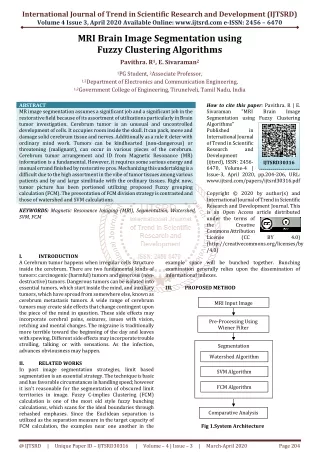
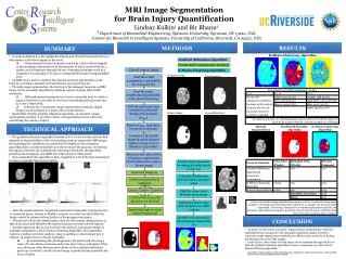
MRI Image Segmentation for Brain Injury Quantification. Lindsay Kulkin 1 and Bir Bhanu 2 1 Department of Biomedical Engineering, Syracuse University, Syracuse, NY 13210, USA 2 Center for Research in Intelligent Systems, University of California, Riverside, CA 92521, USA. METHODS. RESULTS.

E N D
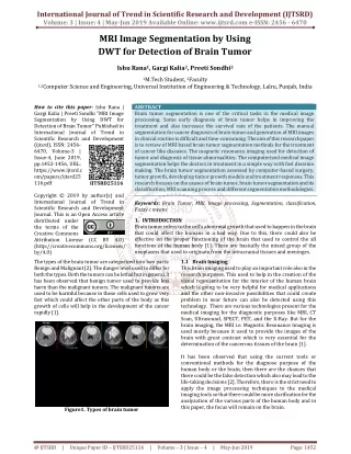
MRI Image Segmentation for Brain Injury Quantification Lindsay Kulkin1 and Bir Bhanu2 1 Department of Biomedical Engineering, Syracuse University, Syracuse, NY 13210, USA 2 Center for Research in Intelligent Systems, University of California, Riverside, CA 92521, USA METHODS RESULTS SUMMARY K-Means Clustering Algorithm • A stroke is defined as a the rapidly developing loss of brain function(s) due to a disturbance in the blood supply to the brain. • Characterized as either Ischemic (caused by a lack of blood supply) or Hemorrhagic (characterized by the presence of extra vascular blood), a stroke can be diagnosed through the use of imaging techniques such as a Computed Tomography (CT) scan or a Magnetic Resonance Imaging (MRI) scan. • An MRI can be used to visualize the internal structure and function of the brain by providing a detailed contrast between several soft tissues. • Through image segmentation, the location of the damaged region in an MRI image can be accurately identified by labeling regions of pixels with similar intensities. • Although manual segmentation is most commonly used to outline a region of interest, it can often be very time consuming and inaccurate due to a user’s subjectivity. • With the use of automatic image segmentation methods, digital images can be analyzed in a faster and accurate manner. • Specifically with the gradient relaxation algorithm, an automatic image segmentation method, it provides a faster and repeatable process while also quantifying the volume of injury. Gradient Relaxation Algorithm Connected Components Analysis Upload original image K-Means Clustering Algorithm Original Image First Iteration Find the initial assignment of probability for each pixel (Pi) Find the mean neighborhood probability of the pixel at hand (qi) Find the maximum kept constant for all pixels during each iteration (ρimax) Second Iteration Binary Image By selecting a k value of 4, the distribution of pixels allows for both the regions of injury to be isolated while also preventing imprecise clustering Based on ρimax, find the ρi constant for all pixels TECHNICAL APPROACH Manual Segmentation Gradient Relaxation Algorithm K-Means Clustering Algorithm Construct a threshold image in which all pixel values converge to zero (black) or 255 (white) • The gradient relaxation algorithm (written in C++) is an iterative process that depends on the probability of the surrounding pixels in a particular MRI image. By examining the consistency of a pixel with its neighbors, the relaxation algorithm relies on pixel probability in order to ensure the accuracy of retaining the region of interest. By automatically selecting a threshold, the algorithm partitions the histogram of an MRI into either black or white pixels. • Once segmented, the algorithm is then reapplied to each individual component in order to quantify the region of injury. Based on the valley of the histogram, segment the first iteration and create a binary image Isolate each component by setting all other pixels to zero (3) Invert the image (1) Calculate the distance between each color pixel value Check every pixel for 8 neighbors 3 Label 0 Label 1 Label 2 Find the mean value between each cluster Continue the loop until all pixels are checked Using an automatic image segmentation process, such as Connected Components analysis or K-Means Clustering algorithm, allows for an image to be more precisely segmented. On average, compared to a manual segmentation analysis, the automatic segmentation techniques produced areas of injured regions .83% less then that of the manual segmentation method. Update the mean value until all pixels are within a cluster Apply label2rgb to create colored regions (2) • After the segmentation is completed, connected components (contiguous sets of connected pixels, written in Matlab) analysis is carried out until either the image cannot be segmented any further or the program terminates. • This process allows the identification of pixels with similar characteristics to that of an injury and identifies the injured regions as several colored regions. • Another approach that is also useful for the analysis of grayscale images of multiple components is the K-Means Clustering Algorithm. This algorithm, which is a method of cluster analysis, aims to partition n observations into k clusters (a number that is chosen manually). • By transforming the 3D image into a 2D matrix and choosing a value of k, the distance between each color pixel value is calculated. Then, once the mean value between each cluster is then updated until all the pixels are clustered, a multi-colored image is produced that quantifies the areas of injury. Complete this process for all connected components (4) CONCLUSION • As shown in the results, automatic segmentations methods have relatively small differences compared to the manually segmented images. However, automatic image segmentation methods are effective and consistent in finding the damage area in the MRI images. • In the future, other forms of brain injury can be analyzed through the use of both the gradient relaxation algorithm/connect components as well as the K-Means Clustering algorithm. 1 2 4 I would like to thank graduate students Benjamin Guan, Angello Pozo and Giovanni DeNina, as well as all the Center for Research in Intelligent Systems (CRIS)