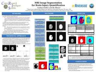
Generative models for automated brain MRI segmentation
Generative models for automated brain MRI segmentation. Koen Van Leemput Athinoula A. Martinos Center for Biomedical Imaging Department of Radiology, MGH Harvard Medical School, USA Computer Science and Artificial Intelligence Laboratory Massachusetts Institute of Technology, USA.


Generative models for automated brain MRI segmentation
E N D
Presentation Transcript
Generative models for automated brain MRI segmentation Koen Van Leemput Athinoula A. Martinos Center for Biomedical Imaging Department of Radiology, MGH Harvard Medical School, USA Computer Science and Artificial Intelligence Laboratory Massachusetts Institute of Technology, USA
Koen Van Leemput DTU Medical Visionday May 27, 2009 MRI of the brain Magnetic resonance imaging: • Harmless • Three dimensional (3-D) • High soft tissue contrast • High spatial resolution • Extremely versatile • Possibly multi-spectral “voxel” Ideal for studying the living human brain
Koen Van Leemput DTU Medical Visionday May 27, 2009 Segmentation of brain MRI • Delineating structures of interest in the images • Segmentation is important: • Basic neuroscience • Uncovering disease mechanisms • Diagnosis, treatment planning, and follow-up • Clinical drug trials • … • Automated computational methods are needed
Koen Van Leemput DTU Medical Visionday May 27, 2009 Overview • Segmentation basics: modeling and inference • Modeling MRI bias fields • Mesh-based brain atlases • Whole-brain segmentation
Koen Van Leemput DTU Medical Visionday May 27, 2009 Overview • Segmentation basics: modeling and inference • Modeling MRI bias fields • Mesh-based brain atlases • Whole-brain segmentation
Koen Van Leemput DTU Medical Visionday May 27, 2009 The problem to be solved MRI image
Koen Van Leemput DTU Medical Visionday May 27, 2009 The problem to be solved MRI image Label image
Koen Van Leemput DTU Medical Visionday May 27, 2009 One solution: generative modeling • Formulate a statistical model of how an MRI image is formed • The model depends on some parameters “labeling model” “imaging model” Label image MRI image
Koen Van Leemput DTU Medical Visionday May 27, 2009 Segmentation = inverse problem MRI image Label image
Koen Van Leemput DTU Medical Visionday May 27, 2009 Segmentation = inverse problem MRI image Label image • Bayesian inference • Start from our statistical model of image formation • Play with the mathematical rules of probability
Koen Van Leemput DTU Medical Visionday May 27, 2009 Bayesian inference Practical approximation Involves two optimizations: • First estimate the optimal model parameters • Then find the optimal segmentation based on those parameter estimates
Koen Van Leemput DTU Medical Visionday May 27, 2009 Example: Gaussian mixture model • The label in each voxel is drawn independently with a probability for tissue type k • Assume a uniform prior for the labeling model parameters “labeling model” “imaging model” Label image MRI image
Koen Van Leemput DTU Medical Visionday May 27, 2009 Example: Gaussian mixture model • The intensity in each voxel is drawn independently from a Gaussian distribution associated with its label • The imaging model parameters are the mean and variance of each Gaussian: • Assume a uniform prior “labeling model” “imaging model” Label image MRI image
Koen Van Leemput DTU Medical Visionday May 27, 2009 Example: Gaussian mixture model three labels • Model parameters are unknown Mean and variance of each Gaussian Relative weight of each Gaussian
Koen Van Leemput DTU Medical Visionday May 27, 2009 Optimization 1: parameter estimation • Given an MRI image to be segmented, what is the MAP parameter estimate ? • Parameter optimization with an Expectation Maximization (EM) algorithm • Repeatedly maximize a lower bound to the objective function • Iterative parameter optimizer using only closed-form parameter updates! current estimate
Koen Van Leemput DTU Medical Visionday May 27, 2009 Optimization 1: parameter estimation
Koen Van Leemput DTU Medical Visionday May 27, 2009 Optimization 1: parameter estimation
Koen Van Leemput DTU Medical Visionday May 27, 2009 Optimization 2: segmentation white matter • Upon completion of the parameter estimation algorithm, assign each voxel to the MAP label CSF gray matter
Koen Van Leemput DTU Medical Visionday May 27, 2009 Overview • Segmentation basics: modeling and inference • Modeling MRI bias fields • Mesh-based brain atlases • Whole-brain segmentation
Koen Van Leemput DTU Medical Visionday May 27, 2009 MRI bias field artifact Intensity inhomogeneities across the image area Imaging artifact in MRI equipment limitations patient-induced electrodynamic interactions MRI data after intensity windowing…
Koen Van Leemput DTU Medical Visionday May 27, 2009 MRI bias field artifact Causes segmentation errors with our segmentation procedure so far…
Koen Van Leemput DTU Medical Visionday May 27, 2009 MRI bias field artifact Causes segmentation errors with our segmentation procedure so far…
Koen Van Leemput DTU Medical Visionday May 27, 2009 Improved imaging model “labeling model” “imaging model” Label image MRI image
Koen Van Leemput DTU Medical Visionday May 27, 2009 Improved imaging model “labeling model” “imaging model” Label image MRI image old model
Koen Van Leemput DTU Medical Visionday May 27, 2009 Improved imaging model “labeling model” “imaging model” Label image MRI image + polynomial bias field model old model
Koen Van Leemput DTU Medical Visionday May 27, 2009 Model parameter estimation • Polynomial coefficients are part of the model parameters • Parameter optimization with a Generalized Expectation Maximization (GEM) algorithm • Repeatedly improve a lower bound to the objective function • Iterative parameter optimizer using only closed-form parameter updates! [Van Leemput et al., IEEE TMI 1999] current estimate
MRI data Estimated bias field Bias-corrected MRI data Koen Van Leemput DTU Medical Visionday May 27, 2009 Example
Koen Van Leemput DTU Medical Visionday May 27, 2009 Example MRI data White matter without bias field model White matter with bias field model Estimated bias field
Koen Van Leemput DTU Medical Visionday May 27, 2009 Example MRI data White matter without bias field model White matter with bias field model Estimated bias field
Koen Van Leemput DTU Medical Visionday May 27, 2009 Overview • Segmentation basics: modeling and inference • Modeling MRI bias fields • Mesh-based brain atlases • Whole-brain segmentation
Koen Van Leemput DTU Medical Visionday May 27, 2009 Improving the labeling model “labeling model” “imaging model” Label image MRI image • So far our labeling model just expresses the relative frequency of occurrence of different labels • Too simplistic for segmenting the brain into 30+ subregions A more realistic labeling model is needed!
Koen Van Leemput DTU Medical Visionday May 27, 2009 Improving the labeling model
Koen Van Leemput DTU Medical Visionday May 27, 2009 Improving the labeling model Try to find the underlying probability distribution Manual segmentations in N individuals (training data)
Koen Van Leemput DTU Medical Visionday May 27, 2009 Modeling the training data (2-D) Triangular mesh representation
Koen Van Leemput DTU Medical Visionday May 27, 2009 Modeling the training data (2-D) “atlas” Assign label probabilities to each mesh node • Flat prior • Label probabilities are linearly interpolated over triangle areas
Koen Van Leemput DTU Medical Visionday May 27, 2009 Modeling the training data (2-D) “atlas” Mesh node positions are sampled from a topology-preserving Markov random field prior warped atlases “knob” that controls the flexibility of the atlas warp
Koen Van Leemput DTU Medical Visionday May 27, 2009 Modeling the training data (2-D) atlas Example segmentations are sampled according to the deformed atlases warped atlases example segmentations
Koen Van Leemput DTU Medical Visionday May 27, 2009 Bayesian inference [Van Leemput, IEEE TMI 2009] Given a collection of manual segmentations • what is the most probable atlas? • what is the most likely value of the parameter controlling the flexibility of the deformations? • what is the most likely mesh representation? Good models explain regularities in the manual segmentations • Automatically yields sparse representations that explicitly avoid overfitting to the training data • cf. Minimum Description Length
Koen Van Leemput DTU Medical Visionday May 27, 2009 Example atlas Derived from manual segmentations of 36 brain substructures in 4 individuals Has average “shape”
Koen Van Leemput DTU Medical Visionday May 27, 2009 Overview • Segmentation basics: modeling and inference • Modeling MRI bias fields • Mesh-based brain atlases • Whole-brain segmentation
Koen Van Leemput DTU Medical Visionday May 27, 2009 Whole-brain segmentation • Tetrahedral mesh-based atlas • The labeling model parameters are the location of the mesh nodes • The prior is the topology-preserving MRF model (penalizes deformations) “labeling model” “imaging model” Label image MRI image
Koen Van Leemput DTU Medical Visionday May 27, 2009 Whole-brain segmentation “labeling model” “imaging model” Label image MRI image + polynomial bias field model Gaussian mixture model
Koen Van Leemput DTU Medical Visionday May 27, 2009 Whole-brain segmentation • Model parameter estimation: • Fully automated segmentation procedure • No need for pre-processing (skull stripping, bias field corr., …) • Automatically adapts to different scanners and acquisition sequences! • Fast! • Improve the imaging model parameters • (Generalized Expectation-Maximization; • closed-form expressions) • Improve the atlas warp • (registration; gradient in analytical form)
Koen Van Leemput DTU Medical Visionday May 27, 2009 Examples (validation under way)
Koen Van Leemput DTU Medical Visionday May 27, 2009 Examples (validation under way)
Koen Van Leemput DTU Medical Visionday May 27, 2009 Examples (validation under way)
Koen Van Leemput DTU Medical Visionday May 27, 2009 Examples (validation under way)
Koen Van Leemput DTU Medical Visionday May 27, 2009 Examples (validation under way)
Koen Van Leemput DTU Medical Visionday May 27, 2009 Examples (validation under way)