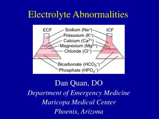
Electrolyte Abnormalities
Electrolyte Abnormalities. Objectives…. Review conditions associated with life threatening electrolyte imbalances Review Hyperkalemia and Hypokalemia causes, diagnosis and treatment Review Hypernatremia and Hyponatremia causes, diagnosis, and treatment


Electrolyte Abnormalities
E N D
Presentation Transcript
Objectives… • Review conditions associated with life threatening electrolyte imbalances • Review Hyperkalemia and Hypokalemia causes, diagnosis and treatment • Review Hypernatremia and Hyponatremia causes, diagnosis, and treatment • Review Hypercalcemia and Hypocalcemia causes, diagnosis, and treatment • Review Hypermagnesemia and Hypomangnesemia causes, diagnosis and treatment • Discuss life threatening acid-base inbalances
Introduction Identified abnormalities most likely represent only the TIP of THE ICEBERG . They are often a small indication of a much larger problem.
Conditions Associated with Electrolyte and Acid Base Problems Signs/Symptoms Acute Conditions Chronic Problems Vomiting Diarrhea/Constipation Confusion, Lethargy, Irritability Weakness / Fatigue Medications Alcohol Abuse Acute Pancreatitis Poor intake Seizures Surgery Peritonitis Overdose Ketoacidosis Respiratory Failure Shock Renal Failure Drug Abuse Metastatic Cancer Immobilization Alcohol Abuse (Chronic) Hyperalimentation Malnutrition Nephrotic Syndrome Diabetes HTN Cirrhosis Heart Failure COPD
PotassiumMOST LIKELY TO BE LIFE THREATENING Potassium is critical for: • Muscular Function • Neurological Function • Minor Changes Affect Conduction and Excitability of the Heart NORMAL RANGE: 3.5 to 5 mEq/L
Hyperkalemia One of the few lethal electrolyte disturbances Endogenous Causes Exogenous Causes MEDICATIONS: K sparing diuretics, ACE inhibitors, NSAIDS, Supplements, PCN, Beta Blockers, Succinylcholine Blood administration (older blood in particular) Diet (RARE) PSEUDOHYPERKALEMIA: WAS YOUR BLOOD SAMPLE HEMOLYZED? • Chronic Renal Failure • Metabolic Acidosis (eg ketoacidosis) • Chemotherapy causing tumor lysis • Rhabdomyolysis • Hemolysis • Hypoaldosteronism
Most common causes of hyperkalemia? Severe, life threatening hyperkalemia is most often caused by kidney failure. Medications are the most common exogenous cause. IN HOSPITAL: Most commonly caused by potassium supplements
DiagnosisSymptoms include: weakness, hypotension, and paresthesia ECG Findings as Potassium increases:
TreatmentStep 1: STOP supplementation and review medications Mild: 5 to 6 mEq/L Severe: 6.5 + with ECG changes FIRST: give 5 to 10 ml of 10% calcium chloride IV over 5 minutes (Calcium will antagonize the toxic effects of potassium on myocardial cell membranes) SECOND: Initiate a rapid shift of K into cells with glucose plus insulin and nebulized albuterol FINALLY: Begin removing excess K from body using methods previously mentioned. Remove excess potassium from the body or cause temporary intracellular shift by: • Diuretics (Lasix 1mg/kg up to 80mg given slow IV) • Resins such as Kayexalate • Dialysis • Glucose plus insulin 10 units R + 25 g glucose IV • Nebulized albuterol (10 to 20mg over 15 minutes)
Hypokalemia: <3.5Life threatening hypokalemia is rare Can cause arrythmia much like hyperkalemia, usually seen as U waves, flat T-waves, and ventricular arrythmias. Often seen with hypomagnesemia Causes most commonly seen are • Poor diet / malnutrition • GI and sweat loss: vomiting, NG suctioning, diarrhea, malabsorption syndromes, heat stroke • Renal losses: Diuretics, aldosteronism • Medications: Aminoglycosides, PCN, Cisplatin, Lithium, L-Dopa, Thallium, Theophyline
Diagnosis 2.6% of hospitalized patients have significant hypokalemia (<3.0). These patients have a 21% mortality rate. • Hospitalized patients often take diuretics. Stress hormones cause decrease in K+. • Mild 3.0-3.5: Often no symptoms • Moderate 2.5-3.0: Generalized weakness, fatigue, constipation, leg cramps. • Severe 2.0-2.5: Muscle breakdown, bowel obstruction • Life threatening < 2.0: Ascending paralysis, impaired respiratory function, unstable arrythmieas
Treatment • BOLUS POTASSIUM DURING CARDIAC ARREST IS NOT ADVISED • Generally, for every 1 mEq/L deficit, the total body deficit is 150 to 400 mEq • pH heavily affects serum potassium. K+ changes 0.3 mEdz for every 0.1 change in pH. Factor this into replacement • Oral or IV - Fast or Slow. In general oral is preferred at 20mEg/h dose. • Maximum IV concentration is 40mEq in 1 Liter of normal saline at a rate of 1000 ml/hr. • MONITOR CONTINUOUSLY FOR IV THERAPY. Do not infuse through central line if the tip of catheter is in the right atrium.
In General • Unlikely to be cause of severe cardiac instability • Most impactful on serum osmolality. Severe increase in sodium = severe increase in serum osmolality. • Abnormalities often reflect total body water abnormalities • Usually levels controlled by the renin-angiotensin-aldosterone system and with ADH. • Sodium is an extracellular ion primarily. ACUTE CHANGES PRODUCE FREE WATER SHIFTS INTO AND OUT OF VASCULAR SPACE • An acute drop in serum sodium will shift water out of vascular space and into interstitial space (can cause cerebral edema) • An acute rise will shift water into the vascular space • Rapid correction of hyponatremia is associated with rhabdomyolysis and cerebral bleeding • As total sodium increases, extracellular fluid increases, resulting in fluid volume overload (CHF, Liver failure, nephrotic syndrome). When total sodium content decreases, ECF volume decreases (volume depletion). Causes orthostatic hypotension, tachycardia, etc.
Hypernatremia Normal range: 135 – 145 mEq/L Causes: Generally 1 of 3 mechanisms Insufficient water intake Loss of water and sodium (but more water than sodium lost) Excess gain of sodium
Hypernatremia Diagnosis and Treatment Diagnosis: Remember that high sodium levels cause shift in fluids from interstitial space to vascular space • Minor Symptoms: Nausea, Vomiting, Fatigue, Irritability • Severe: Confusion, Coma, Seizures, Muscle Weakness, Ataxia Total body water is 50% of lean body weight (Total body water = .5 x kg Treatment of hypernatremia: Correct underlying cause and correct water deficit if present. • Replace water lost is focus. NOT removing excess sodium • Fluid replacement is generally indicated. Use normal saline or equivalent. Water solutions (D5W) reduce concentrations to quickly. • Emergent intervention: 500ml fluid bolus every 20 minutes until stable
HyponatremiaRepresents excess water in relation to sodium Causes: Generally caused by decreased renal excretion of water OR by loss of sodium
Hyponatremia Treatment Use a stepwise approach: • Assess intravascular volume (hypervolemic [edematous states], hypovolemic, or normovolemic) • Treat based on severity of symptoms and volume status • Hypovolemic treat with normal saline • Hypervolemic treat by fluid restriction, diuresis with furosemide • Normal volume status (SIADH) restrict fluid volume and treat underlying cause • About 3% Saline…
CalciumNormal total calcium: 8.5 to 10.5 mg/dL - Normal ionized calcium: 4.2 to 4.8 mg/dL • Most abundant mineral in the body. • Many processes depend on intracellular calcium such as enzymatic reactions, muscle contraction, cardiac contractility, and platelet aggregation. Essential for bone strength and neuromuscular function. • Half of all calcium is bound to albumin • Half of all calcium is biologically active in its ionized form. • Albumin concentration affects serum calcium levels • Ask for lab that includes both if signs of hypocalcemia
HypercalcemiaFairly common (up to 6% of population). Over 90% caused by malignancy or hyperparathyroidism.
Diagnosis“Stones, Bones, Moans and Groans, and Psychologic Overtones” • Stones: Renal lithiasis • Bones: Osteolysis releasing calcium (metastatic disease) • Moans and Groans: Generalized abdominal pain, peptic ulcers • Psychologic Overtones: Apathy, depression, stupor, coma, irritability, hallucinations • Levels greater than 15mg/dL for total calcium may cause cardiac symptoms. AV block can progress to a complete heart block.
If rapid decrease in calcium is needed… … Hemodialysis is the treatment of choice.
HypocalcemiaRelatively rare. Most frequent causes are pancreatitis, Vitamin D deficiency, and medications.
Diagnosis and Treatment Usually asymptomatic. Symptoms usually occur when levels of ionized calcium fall below 2.5 mg/dL • Paresthesia of extremities • Muscle cramps, carpopedal spasms, stridor, tetany, seizures, coma. • Hyperreflexia and positive Chvostek sign (tap over facial nerve in front of ear produces a twitch in eyelid or mouth) • ECG changes include prolonged QT, T wave inversion , bradycardia, AV block and V-tach Treatment: • Give Calcium • Calcium chloride (10%) 5 to 10 mL • Calcium gluconate (10%) 15 to 30 mL IV over 5 minutes • USUALLY HAVE TO CORRECT ABNORMALITIES IN MAGNESIUM AND POTASSIUM
Magnesium • Fourth most common mineral and second most abundant intracellular cation • Necessary for movement of sodium, potassium, and calcium in and out of cells. • Low potassium and low magnesium is a combination for severe arrhythmias • Most magnesium is in bone or bound to albumin, so lab draws for magnesium levels don’t reliably reflect total body magnesium.
HypermagnesemiaCauses and Diagnosis Rare. Almost always associated with renal failure. Symptoms relate to severity: • 3 to 4 mEq/L: Neuromuscular irritability, somnolence, and loss of DTR • 4 to 5: Severe muscle weakness • 5 to 8: Vasodilation and Hypotension • 8 mEq/L or higher: Cardiac conduction problems, neuromuscular paralysis, hypotension, ventilation failure, and cardiac arrest
Treatment • Stop giving Magnesium! • Treated initially with calcium which removes magnesium from serum • May have to support cardiorespiratory function • Magnesium bolus is used frequently in obstetrics. In case of overdose calcium administration may be lifesaving intervention. • 1. Dialysis is ultimate treatment • 2. Stop giving magnesium and support ABCs. • 3. Dilute serum levels with normal saline. • 4. Antagonize effects with calcium. • 5. Remove excess magnesium with diuretic (furosemide 1mg/kg up to 80 mg)
Hypomagnesemia • More common than hypermagnesemia • Interferes with the effects of PTH and results in hypocalcemia. • Can also lead to hypokalemia.
Causes and Diagnosis • Occurs in 11% of all hospitalized patients and 65% of severely ill patients. • Mostly asymptomatic • If any symptoms, usually muscular tremors, altered mentation, vertigo, ataxia • If severe can cause tetany and seizures • Usually associated with alterations in potassium and calcium
Treatment of Hypomagnesemia Treatment depends on severity. Replace cautiously as significant possibility of life threatening hypermagnesemia. • Mild: Oral replacement is preferred. Give Magnesium oxide 400 mg BID • Moderate: give Mag Sulfate 1 – 2 grams over 15 minutes, then 6 grams over 24 hours. MONITOR DTR’s. May take days to see correction. • Severe: Give 2 grams over 15 minutes, then 6 grams every 24 hours for up to 7 days. Check mag levels daily and monitor DTR’s.