Sample Preparation and Mapping
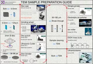
Sample Preparation and Mapping. Steps Cleaning and drying of bulk samples Initial Preparation. Cutting, impregnation, cast-making, fusing Mounting. Stubs, embedded samples, thin sections, grain mounts Polishing. For quantitative analysis Cleaning Mapping Coating Handling and storage.

Sample Preparation and Mapping
E N D
Presentation Transcript
Sample Preparation and Mapping • Steps • Cleaning and drying of bulk samples • Initial Preparation. Cutting, impregnation, cast-making, fusing • Mounting. Stubs, embedded samples, thin sections, grain mounts • Polishing. For quantitative analysis • Cleaning • Mapping • Coating • Handling and storage
Preparing Bulk Samples • Initial Prep: • Remove organic material with oxidizing agent such as K-permanganate or H2O2 • Rinse sediments or soils to remove soluble salts and fines if desired • Carbonate can be removed with HCl • Remove hydrocarbons by soaking in trichlorethane
Drying: • Wet samples must be oven- or freeze-dried before introduction into the instrument. Be aware that temperatures of over 50oC can remove structural water from clay minerals. • Rock samples that have been cut using a saw should be thoroughly dried prior to mounting.
Initial Preparation • Cutting • Large samples must be cut to thin-section size, or cut or broken to a appropriate size for an SEM stub. Typically, diamond-blade circular saw is used. Friable samples may need to first be impregnated. • Impregnating • Friable or porous samples should be vacuum impregnated prior to sample prep. Loose material on the sample surface can cause contamination of the electron column, and unimpregnated porous samples can cause poor vacuum by prolonged outgassing. • Cast-making • SEM visualization of pore structure can be facilitated by vacuum impregnating a sample, followed by dissolution of the sample material using hydrofluoric acid. Topographic details of small fossils can also be facilitated by producing latex cast • Fusing • Bulk analyses of fused rock samples can be made using EMPA techniques. Samples and standards should be fused using the same procedure.
Mounting • SEM Stubs: • SEM samples can be mounted onto Al or C stubs. C can be used for low X-ray background in the case of particulate analysis. Mounting can be done using epoxy, quick-setting glue, double-stick tape (mostly for particulates) or wax. Small grains can be mounted onto double-stick tape or partially dried carbon or silver paint.
Quantitative Analysis Preparation • Embedding, Thin Section, Grain Mounts • These are the most common techniques for preparation of geological samples.
Polishing • Polishing a sample to a flat, unscratched surface is CRITICAL for good quantitative analysis. An uneven, or scratched sample surface can lead to uneven production of x-rays from the sample surface, errors in absorbtion correction, and spurious results.
Polishing • The polishing steps needed for a sample depends on the types of material present. Metallic phases typically polish very easily, whereas minerals with strong cleavage can be problematic. A typical polishing procedure would be as follows: • Grinding the sample to a flat surface, using 100, 260, and 600 mesh grit diamond wheels, or equivalents. • Wash sample in water • Polishing the sample using 30 micron diamond grit (600 mesh) for 3 minutes. • Ultrasonically clean sample in deionized water • Polish the sample using 6 micron diamond grit for 3 minute • Ultrasonically clean sample in deionized water • Polish the sample using 1 micron diamond grit for 3 minutes • Ultrasonically clean sample in deionized water • Polish the sample using 0.5 micron diamond grit for 1 minute • Ultrasonically clean sample in deionized water
Cleaning • Any contaminating material, particularly skin oil, on the sample surface will end up contaminating the column. Also, contaminating material under the conductive coating of a sample can cause the conductive coating that will be placed on the sample to bubble and crack, making the sample impossible to analyze • KEEP THE SAMPLE CLEAN AND OIL FREE!!! • 1. After final polish, clean the sample ultrasonically with deionized water • 2. Wipe the sample surface with petroleum ether. • 3. Following the petroleum ether cleaning, handle the sample as carefully as possible. Glove handling is ideal, but often inconvenient. An alternative is to handle the sample with a kimwipe, or other lint-free papr or cloth. • 4. Blow the sample off with air prior to coating.
Mapping Finding analysis areas on a sample using the optical microscope or electron imaging can be difficult because of the small field of view. The maximum optical field of view for our instrument is 1750 microns (1.75 mm), and the maximum electron image field is around 2500 microns (2 mm). So, having a good sample map, particularly for quantitative analysis, is very important. Sample mapping is much less important for SEM work. Two types of map may be useful, depending on you sample. 1. Macro-map. Map of entire section. Depending on sample type, this may be hand-sketched, made with a slide copier, or with an enlarging xerox machine. 2. Micro-map. Map of areas of analytical interest. This needs to be produced with a camera-equipped microscope, and can be used to locate and document exact analytical spots. A micromap can also be produced using the microprobe. You can also mark areas of interest on your sample, but this must be done on a clean sample surface prior to applying the conductive coat.
Sample Coating Many geological samples are nonconductors of electricity. Therefore, if an uncoated sample is placed in the path of the electron beam, the sample will charge, causing deviation of the electron beam, as well as catastrophic decharging. The sample must be coated with conductive material, such as carbon, gold, or gold-palladium alloy. CARBON: For quantitative analysis and X-ray mapping, carbon is the coat of choice. Because of its low Z, it has a minimal effect on the X-ray spectrum, either in terms of producing X-ray lines or absorbing X-rays. A carbon coat is applied using a vacuum evaporator at pressures of less than 10-4 torr. The ideal coating thickness is 20 nm. GOLD: Gold can be a better choice for SEM coating because of its higher secondary electron yield.