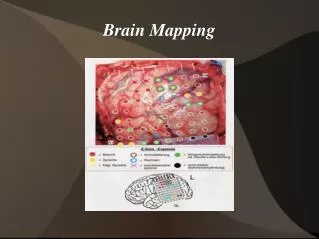
Mapping the Brain
Mapping the Brain. Pages 128-134. Daily Learning Objectives: THE STUDENT WILL . Describe why we call them Brain waves Explain scanning techniques, such as electrode methods Compare and Contrast 2 brain scans. Main room in the house= the Brain. Write Hamlet, discover radium, paper clips

Mapping the Brain
E N D
Presentation Transcript
Mapping the Brain Pages 128-134
Daily Learning Objectives: THE STUDENT WILL • Describe why we call them Brain waves • Explain scanning techniques, such as electrode methods • Compare and Contrast 2 brain scans
Main room in the house= the Brain • Write Hamlet, discover radium, paper clips • How do we study Brain? • Lesion Method- Damaging or removing sections of brain in animals then observing the effects
Electrical and Magnetic Detection • Probed with devices called electrodes • Electrical activity of millions of NEURONS in a region • Measure Brains, activity, damage • Transfers to machine that turns electrical energy into wavy lines on piece of paper • As a result called brain waves
chapter 4 Electroencephalogram A recording of neural activity detected by electrodes
EEG • EEG – electroencephalogram • Brain-wave recording • Standard EEG sounds like outside A.T.&T. park, hear loud noise but not sure what happened • Needle electrodes are more precise- compare and contrast elec. currents • Computer technology is almost always improving so EEG’s analysis is as well
chapter 4 Transcranial magnetic stimulation (TMS) Involves delivering a large current through a wire coil on a person’s head Can be used to Produce motor responses Temporarily inactivate an area of the brain Treat depression
TMS • TMS= transcranial magnetic stimulation • 40,000 times more powerful than Earth’s natural magnetic field • Virtual lesion method • Neurons fire • Produce motor responses- knee jerk, twitch thumb • Important findings- TMS show imagine visual stimulus, the same as seeing a picture
chapter 4 Positron emission tomography Active areas have increased blood flow. Sensors detect radioactivity. Different tasks show distinct activity patterns. A method for analyzing biochemical activity in the brain, using injections of a glucose-like substance containing a radioactive element
Scanning the Brain • PET Scan- 1970’s, Positron-emission tomography • Blood flow, oxygen measures, emotional disorders, active during emotions, activities • Sad memory, math prob. See pretty girl • Nerve cells convert to glucose • SO inject with glucose like substance and substance goes to active brain areas • As a result computer processed picture of biochemical activity on screen
chapter 4 Magnetic resonance imaging (MRI) Magnetic fields align certain ions and compounds. When field is removed, these molecules release energy as radio waves. Computer calculates tissue density from radio waves. Provides clear 3D images Method for studying body and brain tissue
MRI • MRI- magnetic resonance imaging • Allows exploration of inner space • No chemical injection • Magnetic field and radio frequencies • As a result produce vibrations in the nuclei of atoms • Pick up vibrations, turn into signals, computer turns into picture.- Functional MRI
Summary • Electrical magnetic scans • VENN DIAGRAM OF 2 SCANS