CEREBRAL PALSY (CP)
CEREBRAL PALSY (CP). Dr Raj Kumar Yadav Assist. Prof. , PMR MBBS VI Sem. – 25/04/2019. DEFINITION. C linical syndrome has three important criteria - 1. Motor dysfunction. 2. Non progressive brain damage. 3. Affecting an immature developing brain.

CEREBRAL PALSY (CP)
E N D
Presentation Transcript
CEREBRAL PALSY (CP) Dr Raj Kumar Yadav Assist. Prof., PMR MBBS VI Sem. – 25/04/2019
DEFINITION • Clinical syndrome has three important criteria - 1. Motor dysfunction. 2. Non progressive brain damage. 3. Affecting an immature developing brain. • other areas of brain in addition to motor areas • impairments of vision, communication, cognition, mental functions, seizures etc. • during prenatal, during the delivery or postnatal period.
DEFINITION “Cerebral palsy describes a group of permanent disorders of the development of movement and posture, causing activity limitation; those are attributed to nonprogressive disturbances that occurred in the developing foetal or infant brain. The motor disorders of CP are often accompanied by disturbances of sensation, perception, cognition, communication, behaviour, by epilepsy and by secondary musculoskeletal problems”. • Baxet al. 2005 and Rosenbaum
RISK FACTORS A. Prenatal Period: • Pre term (below 36 weeks) • low birth weight (below 2000gms) • TORCH infection • bleeding at third trimester • pre-eclamsic toxaemia • twins and multiple pregnancies
RISK FACTORS • B. Natal Period: • Prolonged labour pains • rupture of placental membrane outside and the time delay from rupture to delivery (if there is delay the child is exposed to infections) • Abnormal presentations like breech • severe hypoxia, bradycardia etc
RISK FACTORS C. Post natal period: • Post encephalitis (both viral and bacterial) • severe hypoxia • Seizures • bleeding disorders, neonatal jaundice and • Traumatic brain injury etc. • The exact time period to label a child with CP due to post natal involvement is variable - 3 - 5 years
PATHOLOGY • Encephalocelewhere a part of the brain is not developed • Microcephaly or Macrocepahly-proliferation of neurons • smooth brain with no gyri known as Lissencepahly, or many small gyri producing Polymicrogyria • Agenises of cortex • Intra ventricular Haemorrhage (IVH) • Cystformations in the cortex • Normal brain structure
CLASSIFIACTION • based on tone changes - 1. SPASTIC TYPE (75%)— • signs of upper motor neuron involvement • Hyperreflexia, Clonus, Extensor Babinski response (abnormal at > 2 years) • Persistent primitive reflexes 2. HYPOTONIC TYPE— • Deep tendon reflexes are weak • unable to maintain the posture • show hyper mobility sometimes.
CLASSIFIACTION 3. DYSKINETIC TYPE— extra pyramidal involvement movement abnormal regulation of tone, defects in postural control and coordination deficits A. Athetoidor slow writhing involuntary movements-in the distal extremities B. Chorea—Abrupt, irregular jerky movements, usually occurring in the head, neck, and extremities C. Choreoathetoid—Combination - Generally large-amplitude involuntary movements. The dominating pattern is the athetoid movement D. Dystonia—A slow rhythmic movement with tone changes generally found in the trunk and extremities, associated with abnormal posturing E. Ataxia—Unsteadiness with uncoordinated movements, often associated with nystagmus, dysmetria, and a wide-based gait
CLASSIFIACTION 4. MIXED TYPE: This includes descriptions from both spastic and dyskinetic classifications. e.g., - spastic athetoid (predominant dyskinetic movement pattern with underlying component of spasticity)
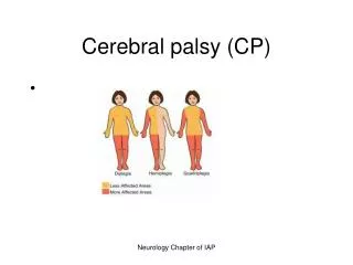
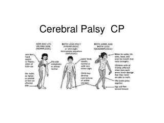
Topographical classification • according to the site of involvement. • Only spastic Neurological type can be classified topographically as other types have generalized body involvement. 1. Spastic quadriplegia: tetraplegia. 2. Spastic triplegia : • classically both lower extremities and one upper extremity. • mild coordination deficits in the uninvolved limb 3. Spastic diplegia: Primarily lower extremity (LE) involvement, mild in coordination problems result in the upper extremities (UE) 4. Spastic monoplegia : Rarely seen, however, has isolated upper or lower extremity involvement and usually a mild clinical presentation 5. Spastic hemiplegia: One side of the body is involved, usually the arm more than the leg.
PRESENTING HISTORY 1. Motor milestones delay. 2. Unable maintain against gravity. 3. Poor hand functions. 4. Preference of one hand before the age of 2 years. 5. Global developmental delay 6. Unable to perform in the school in academic and sports activities. 7. Altered motor performance like bunny hopping, walking with equinus. 8. Poor participation of the child in daily activities like self care, eating etc. 9. Difficulty in handling the child during changing nappies. 10. Sensory issues like poor attention, perceptual disorders, mental impairment.
Epilepsy & MR • 43% of children with CP develop epilepsy, and the risk is increased in those with neuroimaging abnormalities. • Incidence - spastic quadriplegia -50% to 94% - hemiplegia - 30% - diplegiaor ataxic CP - 16% to 27% • Strong correlation of greater intellectual impairment in children who have abnormal EEG findings or epilepsy.
Visual impairment • Common (28%) • Relative risk is increased with the degree of imaging abnormality. • Pathologic disorder of the - anterior afferent sensory visual pathways - ocular structures - cortical vision impairment (CVI) - and disorders of ocular motility.
Hearing impairment • in approximately12% of children with CP. • Risk factors include very low birth weight, kernicterus, neonatal meningitis, severe hypoxic-ischemic insults, and administration of ototoxic medications
Speech and language disorders • Incidence - 38%. • Anarthricor dysarthric speech is caused by oral-motor dysfunction as a result of bilateral corticobulbar dysfunction or lesions to cortical speech-language centers. • Marked motor and speech impairment with relative preservation of intellect is the hallmark of athetoidCP secondary to subcortical basal ganglia lesions.
Voiding dysfunction • 36% of children with CP • In studies where urodynamic evaluation was performed • neurogenic detrusor overactivity with a low bladder capacity was seen in 47% of children with CP • detrusor sphincter dyssynergiawas present in 11%.
Gastrointestinal complications • 27% of children with moderate to severe CP are malnourished. (Quadriplegia > Hemiplegia and Diplegia) - oral-motor dysfunction • Gastroesophagealreflux can be seen because of weakness of the lower esophageal sphincter and can result in emesis and aspiration. • Impaired colonic motility can l/t chronic constipation with possible long-term large bowel megacolon and volvulus, which are preventable with regular bowel evacuation. • Fecal incontinence or defecation distress can occur because of anal sphincter or pelvic muscle incoordination.
Pulmonary complications • 90% of deaths in children with severe CP are caused by pneumonia. • Causes are • pulmonary aspiration • decreased mucociliaryclearance • Suppuration • Kyphoscoliosis • and airway obstruction.
Osteoporosis • In children with CP, bone mineralization can be adversely affected by a combination of factors, including - malnutrition with vitamin D and calcium deficiencies - antiepileptic medications - immobility, and - lack of dynamic loading via muscle forces and standing.
MOTOREXAMINATION: • Passive ROM: • limitation less than 20% • limitation between 20% to 40 % • limitation of more than 40% then surgery shall be needed.
TONE: • NORMAL • SPASTIC - Spasticity will be seen in diplegics, hemiplegics and quadriplegics • FLACCID - hypotonic type, ataxic type • VARIABLE - Dyskinetic • MIXED
STRENGTH: • Smallchild - test in floor and doing functional activities and playing -spontaneous movements. • Grown up children - active movement • Antigravity muscles -responsible for maintaining posture which are called core muscle (Abdominals and Para spinal muscles in particular). • Limb muscles • gross movements of the proximal muscles which are needed for stability and • distal movements in the hands for and fine activities including self care.
POSTURE & BALANCE: • (To be tested without aids splints etc) 1. LYING 2. SITTING 3. KNEELING 4. STANDING 5. WALKING.
CONTRACTURES AND DEFORMITIES • HAND: Thumb in palm, fisted hand, flexion in wrist point towards postural issues of the upper limbs. • LOWER LIMB: Equinous, Knee flexion contracture scissoring
CEREBRAL PALSY GAIT Crouch gait • Hip and knee increased flexion throughout stance with ankle dorsiflexion • Due to hamstring tightness Jump knee gait • Flexion at hip and knee and ankle equinus is characteristic of this gait
GAIT IN CEREBRAL PALSY Stiff knee gait • excess knee extension throughout swing • Has to use circumduction or vaulting • Due to increased rectus femoris activity in swing phase Recurvatum knee • Due to triceps spasticity or hamstrings transfer • Leads to increased knee extension in mid & late stance
SCISSORING GAIT Spasticity of the hip adductors with relative weakness of hip abductors and secondary changes in the hip gives rise to rigidity and excessive adduction of the leg in swing plantar flexion of the ankle increased flexion at the knee
HEMIPLEGIC GAIT • heel strike is missing and patient lands on forefoot • Since hip and knee are kept extended throughout the gait cycle, there is relative limb lengthening and hence circumduction or hip hiking is used for clearance • Toe drag may be present in swing phase • Swing phase is longer on the affected limb • Decreased arm swing on the affected side. • ATAXIC GAIT
COMMUNICATION : • Whether the person with Cerebral Palsy can – 1. Verbalize making sense 2. Communicate with gestures (Speech being unintelligible or inadequate) 3. Communicate with AAC Devices or other gadgets 4. Use some jargon which only the caregivers can understand 5. Cannot communicate
Neonatal reflexes 1. ATNR: Head turned to one side –limbs of same side extended and opposite side flexed 2. STNR: Head flexed in prone position – fore limbs will be flexed and hind limbs will be extended • Head extended in prone position –fore limbs will extend and hind limb will flex 3. Moro: a loud noise or a sudden jerk of the table causes the upper limbs to extend away from the side of the body and then to come together in an embracing pattern. 4. Extensor thrust: when the child is held upright by the armpits, the lower extremities stiffen out straight. 5. Stepping: When held upright, as soon as feet touch a surface, the child places a step forward
INVESTIGATIONS: A. Blood tests: 1. Peripheral smear to look for anaemic status. 2. Serum Vitamin D level in specific cases of malnutrition and risk of fractures before starting therapy. 3. Serum AED (Anti epileptic drugs) to find out for potential toxicity, non responding AED characterized by frequent interruption of rehab program. 4. Thyroid profile if there is family history. 5. Metabolic screening if suspected metabolic involvement
B. Radiology: • X ray pelvis for a child who has • severe scissoring • limitation of passive abduction • less than thirty degrees internal rotation 2. X ray chest 3. X ray of other joints if any severe deformities where serial casting, surgery is planned. 4. X ray spine - spinal curvature
MRI • If there is suspicion of deterioration of function to look for different demyelination diseases. • Location of different MRI findings can tell us the time of insult and the prognosis • To find out structural malformation • As a precursor of metabolic and genetic screening test to be done in cases misdiagnosed as CP. • In some instances for medico legal and disability certifications. • Some lesions in brain can appear later as the brain matures which is not seen earlier MRI.
C. EEG • Seizures • In hemiplegic children who have behavioural issues. D. EMG 1. H reflex studies to confirm spastic patients from benign habitual toe walkers. 2. Hypotonia is seen in some children with DDH and congenital myopathies also. 3. To know the effect of Botulinum toxin A, therapy, medication for reducing spasticity.
E. GENETIC STUDIES: 1. When there is strong family history. 2. Dysmorphic features. 3. When parents want to be clear about planning for another baby. F. BERA G. VEP and Ophthalmic investigations