Download
1 / 26
260 likes | 452 Vues
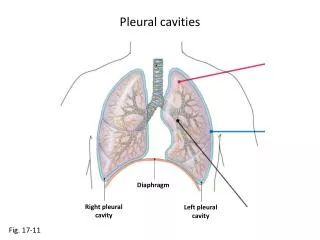
The region between the two pleural cavities that contains the heart and great vessels is called the ______. a. mediastinum b. visceral pericardium c. parietal pericardium d. pericardial cavity. Which layer of pericardium touches the heart?. a. parietal pericardium b. pericardial sac

E N D
The region between the two pleural cavities that contains the heart and great vessels is called the ______. • a. mediastinum • b. visceral pericardium • c. parietal pericardium • d. pericardial cavity
Which layer of pericardium touches the heart? • a. parietal pericardium • b. pericardial sac • c. fibrous pericardium • d. visceral pericardium
Which of the following is true of cardiac muscle tissue? • Cardiac muscle cells are larger than skeletal muscle cells. • Cardiac muscle is not striated. • Cardiac muscle cells have intercalated discs. • Cardiac muscle cells are multinucleate.
Why is the left ventricle more muscular than the right ventricle? • a. It pumps a larger volume of blood. • b. It contracts with force sufficient to push blood through the systemic circuit. • c. The papillary muscles are stronger. • d. Both A and B are correct.
Where is the ANS headquarters for cardiovascular control? • a. cardiac plexus • b. SA and AV nodes • c. medulla oblongata • d. cervical and upper thoracic ganglia
What is the importance of the 100-msec delay at the AV node? • Atria must contract to fill the ventricles with blood. • AV valves must have time to close slowly. • Semilunar valves must have time to close slowly. • Tachycardia results if the delay is absent.
Why is resting HR somewhat slower than the 80–100 bpm set by the SA node? • The AV node slows the heart to an average between its own rate and that of the SA node. • Parasympathetic effects dominate in a resting individual. • Sympathetic fibers release NE to slow heart rate. • Both A and B are correct.
How is cardiac output (CO) calculated? • CO mL/min = (EDV – ESV) × HR • CO mL/min = HR bpm × SV mL/beat • CO mL/min = ESV/EDV • both A and B
During ventricular systole of the cardiac cycle, all of the following would occur EXCEPT_____. • a. rising ventricular blood pressure would exceed aortic pressure • b. all heart valves would be closed • c. atrial diastole would occur as both the atria fill • d. pressure in ventricles would force the semilunar valves closed
When during the cardiac cycle do ventricles contain their maximal amount of blood? What is this quantity called? • at the end of ventricular systole; ESV • at the end of atrial systole; EDV • at the end of ventricular diastole; EDV • both B and C
On an ECG reading, what does the P wave indicate? • a. ventricular contraction • b. an abnormal heart condition • c. atrial depolarization • d. atrial diastole
What event is taking place during the Q–T interval? a. a single cycle of the cardiac cycle b. an action potential c. a single cycle of atrial depolarization and repolarization d. a single cycle of ventricular depolarization and repolarization
What factor could cause an increase in the size of the QRS complex of an electrocardiogram recording? • a. an increase in heart rate • b. a decrease in blood volume • c. a decrease in blood pressure • d. an increase in heart size
What condition contributes to a reduction in the size of the T wave? • a. long-term high fat intake • b. damage to the conduction pathway • c. damage to the AV node • d. coronary ischemia
Why is there no wave corresponding to atrial repolarization on an ECG reading? • It is masked by the QRS complex. • Atrial repolarization produces no electrical effect at all. • It is masked by the P wave. • None of the above is correct.
Which of the following affect(s) the rate of venous return? • a. cardiac output • b. stroke volume • c. heart rate • d. all of the above
How is eversion of the AV valves and backflow of blood into the atria prevented? • a. pressure of blood pushing against the valves • b. contraction of the ventricles • c. closure of the semilunar valves • d. tightening of chordae tendineae and contraction of papillary muscles
Doris was born with a malformed pulmonary valve. How will that affect her circulation? a. Blood will flow more efficiently into her pulmonary trunk. b. Blood will regurgitate into her right atrium. c. Blood will flow back into her right ventricle. d. Deoxygenated blood will continuously pass around her systemic circuit.
Grandpa has developed a radiating pain in his chest upon raking leaves. Which medication might be given to offer prompt relief? a. propranolol, which is a beta 2 blocking medication b. nitroglycerin, which is a vasodilator of coronary vessels c. a fibrinolytic agent to decrease hemostasis d. none of the above
How does damage to the cardioinhibitory center of the medulla affect heart rate? Why? • Heart rate increases; sympathetic dominance. • Heart rate decreases; parasympathetic dominance. • Heart rate remains unchanged; autonomic tone makes delicate adjustments. • Heart rate increases; only the SA node will be controlling heart rate.
Which blood vessels bring blood back into the right atrium? a. foramen ovale, pulmonary trunk, and ductus arteriosus b. superior and inferior venae cavae c. superior and inferior venae cavae and coronary sinus d. aorta, pulmonary trunk, and pulmonary veins
What is the effect of NE binding to adrenergic receptors? • a. increases vasoconstriction • b. decreases heart rate • c. increases heart rate • d. both A and C
Benjamin has an EDV of 120 mL and an ESV of 45 mL, which gives him an SV of 75 mL. What is his ejection fraction? • a. 45% • b. 75% • c. 37.5% • d. 60%
Frank has just run a marathon and his heart is beating extremely rapidly. What happens to the length of diastole and filling time? a. Both increase. b. Both decrease. c. Length of diastole increases and filling time decreases. d. Length of diastole decreases and filling time increases.
Why is ESV lower when you are actively exercising? a. SV decreases and filling time increases. b. EDV is very low and ventricular muscle is stretched very little. c. EDV increases and ventricular muscle produces more forceful contractions ejecting more blood. d. Parasympathetic stimulation causes it.
What is the most important factor in considering cardiac function over time? • a. cardiac output • b. heart rate • c. stroke volume • d. end systolic volume