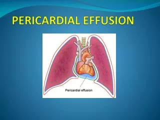
PERICARDIAL EFFUSION
PERICARDIAL EFFUSION. INTRODUCTION. Presence of abnormal amount and/or character of fluid in the pericardial space Can be caused by LOCAL/SYSTEMIC/IDIOPATHIC causes Can be ACUTE or CHRONIC (symptoms)

PERICARDIAL EFFUSION
E N D
Presentation Transcript
INTRODUCTION • Presence of abnormal amount and/or character of fluid in the pericardial space • Can be caused by LOCAL/SYSTEMIC/IDIOPATHIC causes • Can be ACUTE or CHRONIC (symptoms) • Important implications for prognosis (intrathoracic neoplasm), diagnosis (myopercarditis) or both (dissecting of ascentding aorta) • Treatment directed at removal of pericardial fluid and alleviation of the underlying cause
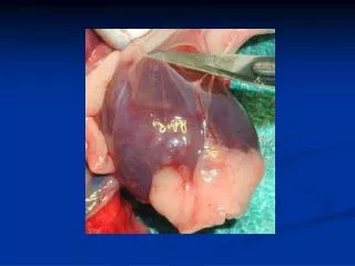
PHYSIOLOGY • Volume of fluid: 15-50 ml. • Essentially and ultrafiltrate of plasma • Total protein generally low. Albumin conc. HIGH. • Contribution of pericardial fluid: end-diastolic pressure (mostly RA,RV)ensure uniform contraction of the myocardium • Acute (80ml) vs. Chronic (up to 2lt).
ETIOLOGY • As a component of any pericardial disorder or 2ry to a systemic disorder: • Acute idiopathic or viral pericaditis • Infectious: Viral, Purulent pericarditis, Tuberculous, HIV • Post MI/post cardiac surgery • Malignancy (lung, breast, hodgkin’s, mesothelioma) • Mediastinal radiation • Autoimmune disease • Dialysis, Ch. Renal failure • Hypothyroidism (myxedema), ovarian hyperstimulation synd. • Drugs: procainamide, isoniazid, hydralazine, anticoagulants.
ETIOLOGY • HEMORRHAGIC PERICARDIAL EFFUSION: • Malignancy (26%) • Trans-catheter interventions and/or pacemaker insertion (18%) • Post-pericardiotomy syndrome (13%) • Complication of MI (free wall rupture, thrombolysis) (11%) • Idiopathic (10%) • Uremic (7%) • Aortic dissection (4%) • Trauma (3%) • Other (8%)
CLINICAL- SYMPTOMS • CVS: chest pain, pericardial pain (relieved by sitting), light headedness, syncope, palpitations • RESP: cough, dyspnea, hoarsness • GI: hiccoughs • NEUR: anxiety, confusion
CLINICAL- SIGNS • CVS:BECK’s triad of tamponade (hypotension, muffled heart sounds, jugular venous distension), pulsus paradoxus, pericardial friction rub, tachycardia, hepatojugular reflux. • RESP: tachypnea, decreased breath sounds, Ewart sign • GI: hepato-splenomegaly • EXTREMITIES: weakened peripheral pulses, edema, cyanosis.
DIAGNOSIS • Suspect when: • All cases of acute pericarditis • Unexplained persistent fever +\- source. Purulent per. • New radiographic cardiomegaly without pul. Congestion. • Isolated left pleural effusion • Hemodynamic deterioration after MI, cardiac surgery, invasive cardiac procedures.
APPRAOCH • Clinically, ECG, X-RAY. • Once pericardial effusion is suspected: • Establish the presence of effusion : clinically ECG, ECHO (sensitive, specific, hemodynamic significance • Assess the hemodynamic impact • Establish the cause
Establish the presence of effusion • According to ACC/AHA/ASE 2003 • Clinically – insensitive and nonspecific. • ECG- low voltage QRS complexes <5mm in all limb leads, <10mm in V1-V6. (tamponade and inflammation); alternans in P and QRS complexes- pathognomonic. • ECHO: sensitive, specific, hemodynamic significance • CT, MRI
Assess hemodynamic impact • Ranges from no significance mild compromisecardiactamponade • Factors determining the degree of hemodynamic compromise: • Volume • Rate of accumulation (acute vs. subacute) • Pericardium is scarred or adherent
Establish the cause of effusion • Often recognized by the clinical setting in which it occurs (cancer, MI, hypothyroidism, renal failure) • Chance of diagnosis rises as the effusion is larger. (15/20% vs. 90%; why? Diag., aggressive approach) • Clinical assess.: size; +/- tamponade; inflammatory signs (chest pain, pericardial friction rub, fever diffused ST elev.) • Lab. Tests: CBC, chemistry+renal function, thyroid, anti dsDNA,complement, chst CT • Pericardiocentesis & biopsy : culture, cytology, PCR.protein,LDH,Glucose,RBC,WBC: do not distinguish exudate from transudate
TREATMENT • CONSIDER: underlying disease, hemodynamic significance, presence of tamponade. • Underlying disease: infectious, malignant, uremic peric. MI, collagen vascular disease. • Cardiac tamponade: volume resuscitation (RA pressure 10-12mmHg). • Pericardial fluid drainage: percutaneous/ pericadiectomy.
Summery • Abnormal amount/character of pericardial fluid • LOCAL/SYSTEMIC/IDIOPATHIC causes • ACUTE vs. CHRONIC • Clinical – not specific. Tamponade. • APPROACH: Clinically, ECG, X-RAY; Establish the presence of effusion ; Assess the hemodynamic impact Establish the cause • TREATMENT: underlying disease, hemodynamic significance, presence of tamponade.