Download
1 / 36
480 likes | 984 Vues
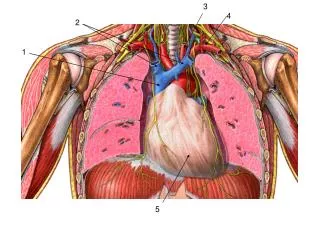
Pericardial Diseases. Visceral – single layer mesothelial cells Parietal- fibrous < 2 mm thick Functions Limits motion Prevents dilatation during volume increase Barrier to infection 15-50 ml serous fluid Well innervated. Acute Pericarditis Etiology. Infectious Viral Bacterial TB

E N D
Pericardial Diseases • Visceral – single layer mesothelial cells • Parietal- fibrous < 2 mm thick • Functions • Limits motion • Prevents dilatation during volume increase • Barrier to infection • 15-50 ml serous fluid • Well innervated
Acute Pericarditis Etiology • Infectious • Viral • Bacterial • TB • Noninfeccious • Post MI (acute and Dresslers) • Uremia • Neoplastic disease • Post radiation • Drug-induced • Connective tissue diseases/autoimmune • traumatic
Infectious • Viral (idiopathic) • Echovirus, coxsackie B • Hepatitis B, influenza, IM, Caricella, mumps • HIV, TB • Bacterial (purulent) • Pneuococcus, staphlococci • fulminant
Pericarditis post- MI • Early <5% patients • Dressler’s 2 weeks – months • Autoimmune • Post-pericardiotomy
Neoplastic • Breast • Lung • Lymphoma • Primary pericardail tumors rare • Hemmorrhagic and large
Radiation • Dose > 4000rads • Local inflammation • Autoimmune • SLE • RA • PSS (40% may develop) • Drugs-lupus like • Hydralazine • Procaimamide • Phenytoin • Methyldopa • Isoniazid • Drugs- not lupus • Minoxidil • Anthracycline antineoplastic agents
Pathogenesis and Pathology • Inflammatory • Vasodilation • Increased vascular permeability • Leukocyte exudation • Pathology • Serous-little cells • Serofibrinous – rough appearance / scarring • common • Purulent – intense inflammation • Hemmorrhagic – TB or malignancy
Clinical • Chest pain • Radiate to back • Sharp and pleuritic • Positional – worse lying back • Fever • Dyspnea due to pleuritic pain
Chest pain in Pericarditis • เจ็บบริเวณหลังต่อกระดูก sternum • เจ็บมากเวลาหายใจ และเวลานอนหงาย • เจ็บน้อยลงเวลาลุกนั่ง และ โน้มตัวไปด้านหน้า
Exam • Friction rub • Diaphragm leaning forward • 1, 2 or 3 components • Ventricular contraction, relaxaltion, atrial contraction • intermittent
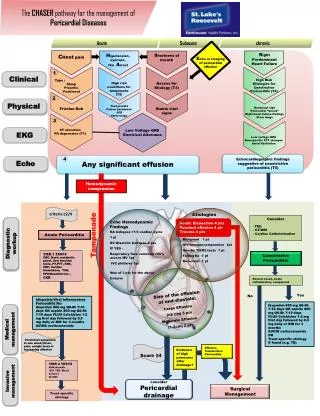
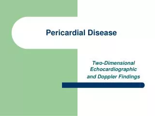
Diagnostic • Clinical history • ECG • Abn in 90% • Diffuse ST elevation • PR depression • Echocardiography • Effusion • PPD • Autoimmune antibodies • Evaluate for malignancy
Treatment • ASA or NSAIDs • Avoid NSAID in MI • Colchicine • Steroids - avoid • May increase reoccurance • TB – Rx TB • Purulent – drainage of fluid + antibiotics • Neoplastic- drainage • Uremic - dialysis
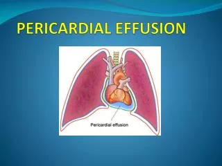
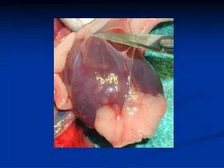
Pericardial Effusion • From any acute pericarditis • Hypothyriodism- increased capillary permeability • CHF- increased hydrostatic pressure • Cirrhosis- decreased plasma oncotic pressure • Chylous effusion- lymphatic obstruction • Aortic Dissection
Effusion Pathophysiology • Pericardium is stiff- PV curve not flat • Above critical volume – rapid increase in pressure • Factors that determine compression • Volume • Rate of accumulation • Pericardial compliance
Clinical • Asymptomatic • Symptoms • CP, dyspnea, dysphagia, hoarseness, hiccups • Tamponade • Exam • Muffled heart sounds • Absence of rub • Ewarts sign-dullness L lung at scapula • atelectasis
Diagnostic studies • CXR - > 250 ml fluid globular cardiomegaly • ECG low voltage and electrical alternans • Echocardiogram most helpful • Identify hemodynamic compromise
Treatment • If known cause- treat that • If unknown- may need pericardiocentesis or pericardial window • Cardiac tamponade is emergency- pericardiocentesis drainage or window
Tamponade • Any cause of effusion may lead to • Diastolic pressures elevate and = pericardial pressure • Impaired LV/RV filling • Increased systemic venous pressure • Decreased stroke volume and C.O. • Shock
Tamponade • Have right side failure with edema and fatigue only if occurs slowly • Key physical findings: • JVD • Hypotension • Small quiet heart • Sinus tachycardia • Pulsus paradoxus- decease in BP > 10 during normal inspiration
Pulsus Paradoxus • Exaggeration of normal • Normally septum moves toward LV with inspiration, with decrease in LV filling • With compression and fixed volume, there is even greater limitation in LV filling and reduced stroke volume • PP also seen in COPD/asthma
Tamponade • Echocardiography • Compression of RV and RA in diastole • Can have localized effuison with localized compression of one chamber (RA,LV) • Effusion post cardiac surgery • Differentiate other causes of low cardiac output • Cardiac catheterization- definitive • Measure pressures- chamber and pericardial equal, and all elevated.
Tamponade- external compression blunts filling throughout cardiac cycle
Pericardial Fluid • Stained and cultured • Cytologic exam • Cell count • Protein level • pp/sp> 0.5 - exudate • LDH level • p LDH/ s LDH > 0.6 - exudate • Adenosine Deaminase level - sensitive and specific for TB
Constrictive Pericarditis • Most common etiology is idiopathic (viral) • Any cause of pericarditis • Post cardiac surgery • Pathology • Organization of fluid, scarring, fusion of pericardial layers, calcification
Constrictive Pericarditis • Impaired diastolic filling of the chambers • Elevated systemic venous pressures • Reduced cardiac output • Dip and plateau curve on catheterization
Constrictive PericarditisClinical • Symptoms • Fatigue, hypotension, tachycardia • JVD, hepatomegaly and ascites, edema • Can confuse with cirrhosis- look for JVD • Exam • Pericardial knock after S2- sudden cessation of ventricular diastolic filling • Kussmaul’s sign- JVD with inspiration • No pulsus paradoxus • Difficult to separate from restrictive cardiomyopathy- may need myocardial biopsy
Normal pericardium < 2 mm (Circulation. 2006;113:1622-1632.)