Download
1 / 1
10 likes | 222 Vues
Comparing Fuzzy Clustering and Finite Gaussian Mixture Model in Brain MRI Jose M. Casillas Diaz. Fuzzy Clustering and FGMM Segmentation Segmentation separates the MR images into its component tissue types. The one we are interested in are: White matter (WM) Grey Matter (GM)

E N D
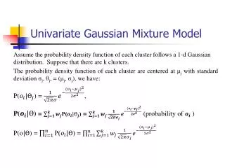
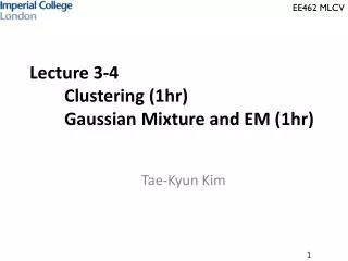
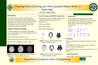
Comparing Fuzzy Clustering and Finite Gaussian Mixture Model in Brain MRI Jose M. Casillas Diaz • Fuzzy Clustering and FGMM Segmentation • Segmentation separates the MR images into its component tissue types. The one we are interested in are: • White matter (WM) • Grey Matter (GM) • Cerebrospinal fluid (CSF) • Introduction • At this time, there are many algorithms for the segmentation of magnetic resonance (MR) images. Fuzzy Clustering and Finite Gaussian Mixture Model (FGMM) are two of them. In this project, we are comparing the accuracy and the precision of the segmentation of the two algorithms. • Magnetic Resonance Imaging • A image technique used to produce high quality cross-sectional images of the internal human body. In this case, the brain. • Uses magnetization and radio waves. • Volumes have magnetization strength of 1.5T and 3T • Each Slice has a thickness of 1mm to 3mm. • Slices can have a size of 512 x 512 or 256 x 256 voxels Methodology By comparing the segmented images with his ground truth (image analyzed by a doctor or expert on the field), we have obtained the confusion matrix which summarizes the results of the classification. (a) (b) (c) • Conclusions • The fuzzy clustering successfully segmented 1.5T and 3T data. • The FGMM have problems segmenting 1.5T data and lower slices that contain the eyes. • The FGMM had better results detectingCSF in some 3T volumes. • Figure 2. Fuzzy clustering segmentation • (a) CSF (b) WM (c) GM (a) (b) (c) • Figure 3. Finite Gaussian segmentation • (a) CSF (b) WM (c) GM Acknowledgements Stacey Franco, Yuhua Gu, Dr. Dmitry Goldgof Figure 1.Axial cross-sectional T1,cerebrospinal fluid and T2 weighted MRI of the brain Department of Computer Science & Engineering