Why do Cells Divide?
Why do Cells Divide?. Cell Size Limits. 1. Replacement of Cells. Humans need to replace old, worn out cells from normal wear and tear. 2. Rate of Diffusion. Remember, the cell membrane allows nutrients (ex. Glucose, oxygen, etc.) in and wastes out (diffusion)

Why do Cells Divide?
E N D
Presentation Transcript
Why do Cells Divide? Cell Size Limits
1. Replacement of Cells • Humans need to replace old, worn out cells from normal wear and tear
2. Rate of Diffusion • Remember, the cell membrane allows nutrients (ex. Glucose, oxygen, etc.) in and wastes out (diffusion) • Diffusion is fast over short distances but slow over long distances • So, b/c of slow rate of diffusion, cells can’t be giants b/c they would starve to death or be poisoned from the build up of wastes!!
Diffusion Animation • Remember diffusion works best over short distances!! • diffusion animation
3. Need enough DNA to support protein needs of cell • Since DNA codes for making protein there needs to be enough to support the protein needs of the cell ex. Think how much protein is in the cell membrane and if the cell size increases than you would need more protein to build it YOU ONLY HAVE SO MUCH DNA AVAILABLE!!
4. Surface Area to Volume Ratio • Need a proper SA:Vol ratio to support the needs of the cell • You need the cell to remain small!! • For example, if the cell size doubles it would need 8x more nutrients to survive and it would create 8x the waste to excrete!! • The vol. increases faster than the surface area of the cell membrane!!
Surface area to volume • surface area to volume
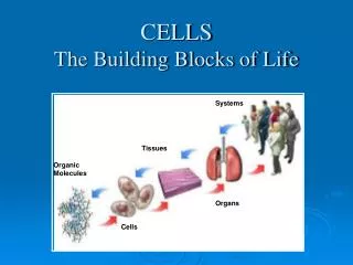
Cell Division Requirements • I) Cell Division • A. Mitosis- nuclear division * • B. Cytokinesis- Division of the cytoplasm
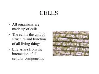
Reminder from cell theory • All cells come from pre-existing cells!! -Cell Division follows this b/c it’s the process where new cells are made from 1 cell • Cell Division makes 2 new daughter cells that are identical to the parent cell
Cell Cycle- Interphase Interphase (Nucleus enclosed in nuclear membrane, nucleolus present, chromosomes appear as chromatin (in other words, chromosomes are not yet visible)) • a) G1 phase- intense cellular activity and growth • b) S phase- DNA synthesis- chromosomes replicate (copy)
Interphase continued • c) G2 phase- spindle fibers are assembled/ centrioles replicate - cell prepares for cell division
Prophase (1st stage) Prophase- 1st stage of actual cell division • a.) Chromosomes condense and shorten, become distinguishable b.) Chromatids attached near center by centromere • c.) Centrioles separate, migrate to the poles • d.) Spindle fibers form between centrioles, asters form • e.) Nuclear membrane disintegrates, nucleolus disappears (in order to separate contents of nucleus, it must disappear)
Prophase Late prophase Early prophase
Metaphase- 2nd stage (shortest) • a.) chromosomes line up in pairs at equator (center of cell) • b.) centromere of each chromosome is attached to a separate spindle fiber
Metaphase diagram Metaphase
Anaphase- 3rd stage • a.) Centromere of each chromosome separates • b.) Separation of the chromatids in each pair • c.) Spindle fibers appear to shorten, pulling the chromatids apart at the centromere (now called chromosomes) • d.) migration of the chromosomes ends with the arrival at the poles and the formation of clusters
Anaphase diagram Anaphase
Telophase – 4th stage • a.) Cleavage furrow forms b/w 2 cells and spindle fibers disappear • b.) Nuclear membrane forms around each set of chromosomes (forms 2 new nuclei) • c.) Chromosomes uncoil to form chromatin net (getting ready for interphase) • d.) Nucleoli reorganized
Telophase Diagram Telophase
Cytokinesis • Cytokinesis- division of the cytoplasm • In animal cells- cell membrane pinches together, furrow forms along the equator. • -In plant cells- cell plate forms in the middle of the dividing cell