Central Nervous System and Peripheral Nervous System
830 likes | 1.22k Vues
Central Nervous System and Peripheral Nervous System. Anatomy and Physiology Assessment Diagnostic Tests. Objectives. Name the two structural divisions of the nervous system and give the functions of each List the parts of the neuron and describe the function of each part

Central Nervous System and Peripheral Nervous System
E N D
Presentation Transcript
Central Nervous System andPeripheral Nervous System Anatomy and Physiology Assessment Diagnostic Tests.
Objectives • Name the two structural divisions of the nervous system and give the functions of each • List the parts of the neuron and describe the function of each part • Explain the anatomic location and functions of the cerebrum, brainstem, cerebellum, spinal cord, peripheral nerves, and cerebrospinal fluid
Objectives • Discuss the parts of the peripheral nervous system and how the system works with the central nervous system • List the 12 cranial nerves and the areas they serve
Objectives • Identify the significant subjective and objective data related to the nervous system that should be obtained from a patient during assessment • Differentiate between normal and common abnormal findings • List the physiologic changes that occur in the nervous system with aging • List common laboratory and diagnostic examinations for evaluation of neurologic disorders
General Overview • The nervous system is responsible for communication and control within the body • It interprets or processes information received and sends it to the appropriate area of the brain or spinal cord where the response is generated • It works in conjunction with the endocrine system to maintain homeostasis
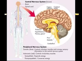
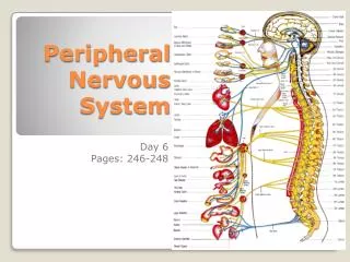
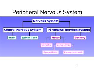
Overview of Anatomy and Physiology • Structural divisions • Central nervous system (CNS) • Brain and spinal cord – interpret incoming sensory information, and issues instructions based on past experience • Peripheral nervous system • Somatic nervous system (conscious control) – sends messages from the CNS to skeletal muscles (voluntary muscles)
Overview of Anatomy and Physiology • Autonomic nervous system (“involuntary NS – without conscious control). It transmits messages from CNS to the smooth muscle, cardiac muscle, and some glands. • Divided into: • Sympathetic • Parasympathetic • Enteric
Overview of Anatomy and Physiology • Cells of the nervous system • Neuron – 2 categories: • Neuron – transmitter cells; carry messages from the brain and spinal cord • Glial or neuroglial cells- supports and protects neurons while producing CSF • Neuron = the basic unit of the nervous system • Composed of 3 parts: • Cell body • Axon • dendrites
Axons and Dendrites Branch off the main cell body Axons conduct impulses away from the cell body Dendrites convey impulses toward the cell body
Overview of Anatomy and Physiology • Neuromuscular junction • The area of contact between the ends of a large myelinated nerve fiber and a fiber of skeletal muscle • SYNAPSE: the “gap” or space between each neuron. Nerve impulses are transmitted across this “gap” or synapse by the action of “neurotransmitters”. • Neurotransmitters • chemicals • Act to make sure that the neurological impulse passess from the nerve to the muscle
Neurotransmitters • Functions • Acetylcholine: impulse transmission • Norepinephrine: Maintaining arousal (from deep sleep), dreaming, mood regulation • Dopamine: motor function; emotional responses • Serotonin: induces sleep; controls temp.; mood control; affects sensory perception
Overview of Anatomy and Physiology • Neuron coverings • Many neurons have a white, waxy, fatty material called “myelin” • Myelin increases the rate of transmission of impulses and protects and insulates the fibers • Axons leaving the CNS are wrapped in layers of myelin with indentations called the nodes of Ranvier • These nodes further increase the rate of transmission because the impulse can jump from node to node
Overview of Anatomy and Physiology • In the peripheral nervous system, the myelin is protected by Schwann cells • Outer layer of Schwann cells protected by another layer called “neurilemma” which helps to regenerate injured axons Regeneration can only happen in the PNS
Cerebrospinal Fluid (CSF) Bathes the structures of the CNS Composed of water, glucose, sodium chloride, and protein Acts as a shock absorber for the brain and spinal cord
Central Nervous System • One of two divisions of the nervous system • Composed of the brain and spinal cord • Functions somewhat like a computer but more complex • Cranium protects the brain • Vertebrae protect the spinal cord
Overview of Anatomy and Physiology • Central nervous system • Brain • Cerebrum • Diencephalon • Cerebellum • Brain stem • Midbrain; pons; medulla oblongata; coverings of the brain and spinal cord; ventricles • Spinal cord
Central Nervous System • Brain • Specialized cells (in the brain’s mass of convoluted soft, gray or white tissue) coordinate and regulate the functions of the CNS • One of the largest organs in the body • Approx 3 lbs • 4 parts: cerebrum, diencephalon, cerebellum, brainstem
Cerebrum • Largest part of the brain • Divided into: left and right hemispheres Each hemisphere controls the opposite side of the body: the right hemisphere controls the left side of the body, and the left hemisphere controls the right side of the body • Outer portion = cerebral cortex (“gray” matter) • Arranged in folds called gyri (convolutions) • Grooves called sulci (fissures)
Cerebrum Largest part of the brain Divides the two hemispheres into 4 lobes named for the bones over them: frontal, parietal, temporal, and occipital lobes Corpus Callosum = the connecting bridge between the two hemispheres
Cerebrum • Function: controls initiation of movement on the opposite side of the body • Specific areas of the cerebral cortex are associated with specific functions *NOTE: see box 14-1 p. 654
(From Thibodeau, G.A., Patton, K.T. [1987]. Anatomy and physiology. St. Louis: Mosby.) Sagittal section of the brain (note position of midbrain).
Diencephalon • The “interbrain”: lies beneath the cerebrum • Contains the thalamus and the hypothalamus. • Thalamus: serves as a relay station for some sensory impulses while interpreting other sensory messages such as pain, light touch, and pressure
Diencephalon • Hypothalamus: • Vital role in temperature, fluid balance, appetite, and emotions such as fear, pleasure, and pain. • Sympathetic and parasympathetic nervous systems are under the control of the hypothalamus • Pituitary gland also under control of hypothalamus
Cerebellum (“little brain”) Lies posterior and inferior to the cerebrum The 2nd largest portion of the brain Also contains 2 hemispheres Uses information received from the cerebrum, muscles, joints, and inner ear to coordinate movement, balance, and posture
Cerebellum (“little brain”) • Unlike the cerebrum, the right side of the cerebellum controls the right side of the body, and the left side of the cerebellum controls the left side of the body
Brainstem • Located at the base of the brain • Contains the midbrain, pons, and medulla oblongata • These structures connect the spinal cord and the cerebrum • Carries all nerve fibers between the spinal cord and cerebrum
(From Thibodeau, G.A., Patton, K.T. [1987]. Anatomy and physiology. St. Louis: Mosby.) Sagittal section of the brain (note position of midbrain).
Brainstem • Midbrain: responsible for motor movement, relay of impulses, auditory and visual reflexes • The origin of cranial nerves III and IV • Pons (“bridge”): responsible for sending impulses to the structures superior and inferior to it • Contains a respiratory center • The origin of cranial nerves V through VIII
Brainstem • Medulla Oblongata: controls heartbeat, rhythm of breathing, swallowing, coughing, sneezing, vomiting , hiccups
Protective Structure of the Brain • Meninges- three layers that surround both the brain and spinal cord • Dura mater – outermost membrane • Arachnoid membrane – 2nd layer • Pia mater – innermost membrane; provides oxygen and nourishment to the nervous tissue • These layers bathe the spinal cord and brain in CSF fluid
Ventricles • 4 spaces or cavities located in the brain • CSF flows into the subarachnoid spaces around the brain and spinal cord and cushions them • CSF contains protein, glucose, urea, salts • Also contains substances that form a protective barrier (the blood-brain barrier) that prevents harmful substances from entering the brain or spinal cord
Spinal Cord • 17-18 inches • Extends from the brainstem to the 2nd lumbar vertebra • 31 pair of spinal nerve roots exit the spinal cord • 2 main functions: • Conducting impulses to and from the brain • Center for reflex actions
Disks Vertebrae separated by disks which serve as shock absorbersfor the vertebral column Composed of Anulusfibrosus: ring of tissue; encircles nucleus pulposus Nucleus pulposus: saclike structure with a gelatinous filling that has a high water content As we age, nucleus pulposus loses much of its water; less effective as a shock absorber
Pyramids and Pyramidal Tracts • Pyramidal Tracts carry motor information from the CNS neurons to the PNS neurons • In the medulla of the brainstem, information from one side of the brain “crosses over” and goes down the pyramidal tracts to affect the other side of the body. • The area where the “crossing over” occurs is the Pyramids
Spinal Cord • Reflexive Action • Sensory neuron sends information to the cord • A central neuron (located within the cord) interprets the impulse • Motor neuron sends the message back to the organ or muscle involved A message is sent, interpreted and acted upon without having traveled to the brain • E.g. knee jerk
Ganglia and Nuclei • Ganglia are information “hubs” of the PNS • Eg. Dorsal root ganglia contain the cell bodies of sensory neurons • Nuclei are information “hubs” of the CNS • Eg. Basal Ganglia in the brain contains neurons connecting the cerebral cortex, thalamus, and brainstem. (incorrectly named; should be “Basal Nuclei)
Overview of Anatomy and Physiology • Peripheral nervous system • Spinal nerves (31 pairs) • Mixed nerves: transmit sensory information to the spinal cord through afferent neurons and • motor information sent to the CNS to the various areas of the body through efferent neurons • Named according to the corresponding vertebra • E.g. C1, C2, T12, L3