Download
1 / 74
750 likes | 981 Vues
Nervous System Central and Peripheral. MED 164. William Budd Medical Careers Institute. Objectives. Functions of nervous system Homeostatic mechanisms Organization of nervous system and components of each branch Autonomic nervous system effects Structure of a neuron

E N D
Nervous SystemCentral and Peripheral MED 164 William Budd Medical Careers Institute
Objectives • Functions of nervous system • Homeostatic mechanisms • Organization of nervous system and components of each branch • Autonomic nervous system effects • Structure of a neuron • Classifications of neurons • Types of neuroglia and functions • Neuronal conduction
Objectives • Structure and function of a synapse • Structure and function of organs in nervous system • Common pathologies of nervous system
Functions of Nervous System • Control and coordination of all body systems and relates an individual to their environment • Homeostatic regulation • Seat of intellect and reasoning
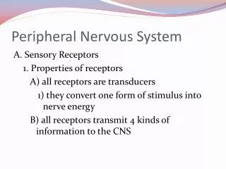
Homeostatic Control • Sensory Input • Receptors monitor changes (stimuli) inside and outside body and information is sent to CNS by an afferent pathway • Integration and interpretation • Sensory information is processed and interpreted and a response decision is made and sent out • Motor Output • Response is sent out by efferent pathway to an effector organ
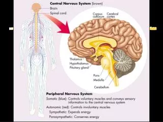
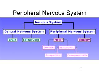
Nervous System Central Nervous System Peripheral Nervous System Sensory (Afferent) Motor (Efferent) • Brain • Spinal Cord Somatic Autonomic Parasympathetic Sympathetic
Central Nervous System • Contains the brain and spinal cord • Seat of integration and interpretation
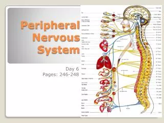
Peripheral Nervous System • Consists of nervous system tissue that is outside of CNS • Primarily functions to communicate between organs and CNS • Contains two types of nerves • Spinal • 31 pairs of nerves that connect to spinal cord and transmit signal to and from spinal cord • Cranial • 12 pairs of nerves that connect to the brain and transmit information to and from brain
Peripheral Nervous System • Two functional subdivisions • Sensory (Afferent) • Transmits information from receptors in tissues and organs to the CNS • Motor (Efferent) • Transmits information from CNS to effector organs such as muscles and glands • Contains two subdivisions; somatic and autonomic
Peripheral Nervous System • Somatic nervous system contains motor fibers that transmit impulses from CNS to skeletal muscles • Commonly called the voluntary nervous system
Peripheral Nervous System • Autonomic Nervous System consists of motor fibers that innervate smooth muscle, cardiac muscle, and endocrine glands • Commonly called the involuntary nervous system • Contains two functional subdivisions • Sympathetic (fight or flight) • Innervated by thoracic and lumbar ganglia of spinal nerves • Parasympathetic (Resting and digesting or feed and breed) • Innervated by vagus nerve and nerves of pelvis
Nervous Tissue • Human body contains 100 billion neurons • Neurons are specialized cells designed to receive information from other neurons or sensory receptors and transmit to other neurons, muscle cells, and/or glands • Neurons are unable to undergo mitosis • Highly active cells that are very sensitive to glucose and oxygen levels
Anatomy of a Neuron • Soma – cell body of a neuron • Somas are located in gray matter of CNS • Brain • Cerebral cortex is composed of neuronal somas • Nuclei are somas located deep in brain tissue; surrounded by white matter • Spinal cord • Cell somas are located deep to white matter in spinal cord • Groups of cell bodies in PNS are called ganglia
Anatomy of a Neuron • Cytoplasm • Contains numerous mitochondria • Neurons utilize large amounts of glucose and oxygem • Contains large amounts of rough ER • Sometimes called Nissi bodies
Anatomy of a Neuron • Neurons contain two types of cellular projections • Dendrites are short, branched projections that conduct impulses towards the soma • Neurons often contain many dendrites • Axons are long, singular processes that conduct impulses away from soma
Anatomy of an Axon • Axonal hillock – cone shaped region where soma tapers into axon • Telodendria – terminal branches of axon • Axon terminals – button shaped endings of telodendria • Sometimes called synaptic terminals, or synaptic knobs • Axons do not contain ribosomes, Rough ER, or Golgi bodies • Do contain an extensive cytoskeleton involved in bi-directional transport
Function of Axon • Axonal terminals are secretory component of axon • Neurotransmitters are stored and released into synapse from axonal terminal
Groups of axons • Axons are often found bundled together with other axons that share a common origin and destination • In CNS, bundles of axons are called tracts • In PNS, bundles of axons are called a nerve • Bundles of axons are supported by glial cells and in PNS protected by connective tissue
Functional Classification of Neurons • Two systems are used to classify neurons • Based on direction of impulse • Based on structure that is innervated and direction of impulse
Classification based on direction • Sensory Neurons (Afferent) • Impulses travel from sensory receptors to CNS • Motor Neurons (Efferent) • Impulses travel from CNS to effector organs (muscles or glands) • Interneurons (Association) • Connect motor and sensory neurons or between interneurons, only in CNS • Accounts for 99% of neurons in CNS
Classification based on tissue innervation • Four functional types of neurons found in body • Somatic neurons innervate muscles involved in voluntary activities • General somatic afferent • General somatic efferent • Visceral neurons innervate muscles involved in involuntary activities • General visceral afferent • General visceral efferent
Supporting Cells • CNS does not contain any connective tissue • Collectively called neuroglia (nerve glue) • 4 types in CNS • 2 types in PNS • Do not generate or transmit electrical impulses • Possess capability to divide and multiply
CNS Neuroglia • Microglia – Macrophages that become phagocytic upon injury to the CNS • Possibly involved in ALS and MDA • Astrocytes – Star shaped cells that are found between soma of neuron and blood vessel capillaries • Provide structural support, repair mechanisms, and metabolic functions that assist neurons
CNS Neuroglia • Oligodendrocyte forms myelin around CNS axons • Ependymal Cells – Cells that line cavities in brain and spinal cord • Responsible for secretion and movement of cerebrospinal fluid
PNS Neuroglia • Schwann cells – Responsible for formation of myelin on peripheral nervous system neurons • Satelite cells – Surround and support neuron cell bodies within ganglia
Myelinated Neurons • Myelinated axons have a multilayered wrapping of the plasma membrane of oligodendrocytes or schwann cells • White matter is a collection of myelinated axons in brain and spinal cord • Functions of myelination is to insulate axon and speed impulse of nerve conduction • Nodes of Ranvier are gaps in myelin sheath
Pathologies Caused by demyleination • Guillein-Barre Syndrome • Multiple Sclerosis • Devic’s Disease (Causes blindness) • Progressive MulitfocalLeukoencephalopathy • Transverse Myelitis
Cellular Conduction • Differences in electrical potentials across cell membrane creates a polarization • At rest, neurons and muscles contain large amounts of potassium inside of cell and sodium outside of cell • When stimulated, a sodium channel opens and sodium travels down its concentration gradient and electrical difference decreases (voltage moves closer to zero) • When voltage reaches a certain point, potassium channels open and potassium leaves the cell
Cellular Conduction • The signal continues down through the neuron • Re-polarization begins when potassium starts to leave the cell and concludes with the activation of a sodium/potassium pump that is utilized to return the cell to its starting position • Process is accomplished within a few milliseconds and occurs millions of times per minute
Types of stimulus • Stimuli can result in one of two types of potentials to occur • Excitatory potentials occur when the stimulus causes the inside of the cell to become less negative (depolarized • Inhibitory potentials occur when the stimulus causes the inside of the cell to become more negative (hyperpolarized)
How do they occur? • Depolarization occurs when sodium channels open and sodium comes into cell • Hyperpolarization occurs when potassium channels open and potassium leaves the cell or chloride channels open and chloride enters the cell
Polarization Muscle Cell - 70mV 30 mV Na+ Na+ Na+ Na+ Na+ 130 mV Na+ Na+ Na+ Na+ K+ K+ K+ K+ K+ K+
Synapses • Neuron does not touch muscles and in only very rare circumstances does it connect directly to other neurons • Two types of synapses • Electrical • Rare; occurs when two neurons connect directly together and allows current to flow directly from one neuron to another • Chemical • Most common; neuron does not connect to anything but is seperated by a synaptic cleft that must be crossed with a chemical neurotransmitter
Synaptic Physiology • Action potential travels down axon and causes calcium channels in pre-synaptic terminal to open and calcium rushes into neuron • Calcium causes vesicles containing neurotransmitter to fuse with cell membrane and neurotransmitter is exocytized into synaptic cleft • Neurotransmitter binds reversibly to specific receptors on post-synaptic membrane
4. Neurotransmitter binding causes an ion channel in post-synaptic membrane to open • As long as neurotransmitter is bound to receptor, channel remains open. • Neurotransmitter must be removed from synapse • Degraded by specific enzymes (acetylcholine) • Removed by astrocytes (Norepinephrine) • Re-uptook back to presynaptic neuron (Serotonin)
Chemicals that affect synapse • Botulinum toxin • Black widow venom • Nicotine • Sarin gas • Organophosphate insecticides • Medications
Conditions affected by synaptic physiology • Depression • Parkinson’s disease • Myasthenia gravis • Drug addiction and alcohol
Meninges • Protective connective tissue covering of the brain and spinal cord • Contains three layers • Dura mater (Tough mother) – Outermost layer of meninges • Composed of two layers of tough dense connective tissue • Archanoid mater (Spider mother) – Loose middle covering that contains weblike extensions that connect to the pia mater
Meninges • Pia mater (Tender mother) – Delicate inner layer of meninges that contains many small blood vessels
Meningeal Spaces • Epidural space – Located between the dura mater and the skull/ vertebral column • In skull contains arteries • In spinal cord contains fat and veins • Subdural space – Potential space between dura mater and arachanoid mater • Subarchanoid space – Space between arachanoid mater and pia mater • Filled with cerebrospinal fluid
Ventricles • Fluid filled cavities within the brain • Lateral ventricles – Two ventricles; one on each side of the brain in the cerebrum • Ventricular foramen – Channel that connects the lateral ventricles to third ventricle • Third ventricle – Inferior to lateral ventricles and enclosed by diencephalon
Ventricles • Fourth ventricle – Dorsal to pons, ventral to cerebellum, communicates with subarchanoid space and continuous with central canal of spinal cord
Cerebrospinal Fluid • Fluid surrounds brain and spinal cord • Cushions brain and gives buoyancy to CNS • Effectively reduces weight of brain • Protects CNS from trauma • Aids in nourishment of nerual tissue • Produced by choroid plexus in ventricles • Choroid plexus is network of capillaries in pia mater • Blood brain barrier seperates substances in blood from brain tissue
Flow of CSF in CNS • CSF is made in lateral ventricles and flows to the third ventricle through the ventricular foramen • Flows through the cerebral aquaduct to the fourth ventricle • Flows to the central canal of spinal cord and down the spine • Fluid moves into the subarchanoid space of the meninges • Reabsorbed in the sagital sinus of the brain by arachanoidvilli
CSF and Diagnosis • CSF can be used to diagnose pathologies of the meninges and brain or used to treat some conditions • Drawn out through a procedure called a lumbar puncture