Download
1 / 88
890 likes | 1.11k Vues
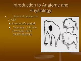
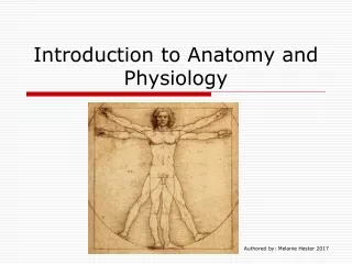
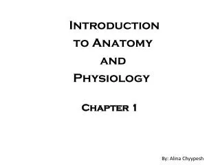
Introduction to Anatomy and Physiology Chapter 1. By: Alina Chyypesh. Answer #2. Give the term, definition and example . Answer # 3. Where is this arrow pointing to? . Answer # 4. What is the name of the body organ system and what it’s functions?. Answer # 5.

E N D
Introduction to Anatomy and Physiology Chapter 1 By: Alina Chyypesh
Answer #2 Give the term, definition and example
Answer # 3 Where is this arrow pointing to?
Answer # 4 What is the name of the body organ system and what it’s functions?
Answer # 5 Explain what is happening in the picture
Slide 6 What is the name of this system and what is the function?
Slide 7 How does the homeostatic control system works?
Answer #8 What are the parts called that are in red?
What are the 3 planes? Answer #9
Answer #10 What is the name of this and give an example?
Answer #11 What is the name and give an example?
Answers • Ex: My skin is superficial to my skeletal muscles. • Lumbar • Muscular System: Allows manipulation of the environment, locomotion, and facial expression. Maintains posture, and produces heat. • This is integumentary system that protects the body as a whole from the outside environment. When you eat something it goes to digestive system which gets broken down into small pieces for the nutrients to be able to be absorbed and it also takes in oxygen which is the repertory system then it is send to the whole body cells. The elimination of metabolic waste goes to the urinary and respiratory system. • Lymphatic System/Immunity: peaks the fluid from blood vessels and returns it back; disposes of debris in the lymphatic stream; houses white blood cells which is involved in immunity, and the respond that it attacks the outside substances within body
Answers • Imbalance variable stimulus: produces change in variable the change is found by the receptor Receptor the information the was found is sent through afferent pathway to Control Center the output: the information is sent through efferent pathway to Effector the response changes the stimulus and returns the variable to homeostasis 8. Superior mediastinum Pleural cavity Ventral body cavity Pericardial cavity Abdominal cavity Pelvic cavity
Answers • Median (midsagittal) plane: cuts half and half top to down Transverse plane: where is stomach and cuts it in half Frontal Plane: cuts in half the back and front • Deep (internal): it is away from the body surface: more inside Ex: The heart is deep to the skin • Ventral (anterior): Toward or at the back of the body; in front of Ex: The ribs are anterior to the spine
Tissues: The Living Fabric Chapter 4
Answer #16 What is the name of this tissue?
Answer #17 What is the name of this tissue and where is it located and what does it do?
Answer #18 What is the name of the tissue and where can you find this type of tissue?
Answer #19 Where is the Nucleus, Rough ER, and Golge apparatus?
Answer #20 What is the name of the tissue and give a description on what you see on the picture.
Answer #21 What is the name of the tissue and where can you find it?
Answer #22 Where is the parietal pleura and visceral pleura?
Where is the parietal peritoneum and visceral peritoneum? Answer # 23
Answers 16. Simple columnar epithelium: absorption, in some parts of uterus 17. Stratified squamous epithelium: protects underlying tissues in areas subjected to abrasion, mouth 18. Simple squamous epithelium: air sacs of lungs, lymphatic vessels 19. Nucleus: a purple circle, Rough ER: blue tubes, Golgi apparatus: green 20. Areolar connective tissue: has 3 fibers, cells, fibroblast, macrophages, mast cells and white blood cells 21. Adipose: under skin, around kidneys and eyeballs 22. Parietal pleura: against body wall and covering lungs, Visceral pleura: covering organ and covering the lungs 23. Parietal peritoneum: against body wall lining abdominal/pelvic cavities, visceral peritoneum: covering organ lining abdominal/pelvis cavities
Answers 24. Nervous tissue: located in the brain, spinal cord and nerves 25. Smooth muscle: function: propels substances or objects along internal passageways, involuntary control
Answer #29 What is this bone made out of?
Tell what you see in this picture Answer #30
Answer #31 What is happening in this picture?
Name the parts that are pointing too Answer #32 3 1 2
Name the parts of the arm bone that the arrow is pointing Answer #33 1 2 3 4
Name the bone that the arrow is pointing and the name Answer #34 2 1
What is the spongy bone made out of? Answer # 35
That are the false and true ribs and how many are there of them? Answer #36 1 2
What is the name of what the arrow is pointing too? Answer #37
What is the name that is circled? Answer #39
Answers • 29. Spongy bone, compact bone, articulate cartilage • Lamella, the yellow: osteocyte, artery, vein, nerve fiber • Forming collar, then cavitation of the hyaline cartilage within the cartilage model, then invasion of internal cavities, then formation of the medullary cavity as ossification continues, then the last one ossification of the epiphyses • 1. supraspinous fossa, 2: infraspinous fossa, 3: Acromion • 1. Lesser tubercle, 2: Deltoid tuberosity, 3: Capitulum, 4: Trochlea • 1:styloid process of ulna, bone: ulna, 2: styloid process of radius, bone: radius • The spongy bone is made out of trabecular • 1: true ribs(1-7), 2: false ribs (8-12) • Collagen fibers • Carpals (wrist)
Name the parts that the arrow is pointing to? Answer #41 1 3 2
What is this movement called? Answer #42
What is the name of this movement? Answer #43
What is the structural type and functional type, and movement allowed? Answer #44
Give the joint, name, stuctural type and tell if has movement Answer # 45
Give the joint, name, structural type and tell if has movement Answer #46
What is the name of the movements that the arrows are pointing to? Answer #47 1 2 2
What is the joint, name, structural type and tell if has movement? Answer #48
From the knee joint: what are the name of the parts that the arrows are pointing too? Answer #50 1 2 3