Download
1 / 23
230 likes | 358 Vues
Helical CT Diagnosis of PTE. Bill Lee. Pulmonary Thromboembolism: Spectrum of Findings on CT Creaves S, et. al. American Journal of Roentgenology 1995;165:1359-1363. PTE. Filling defect in pulm. art. post-contrast Widenening, blunting, abrupt termination of arteries

E N D
Helical CT Diagnosis of PTE Bill Lee
Pulmonary Thromboembolism: Spectrum of Findings on CTCreaves S, et. al.American Journal of Roentgenology1995;165:1359-1363
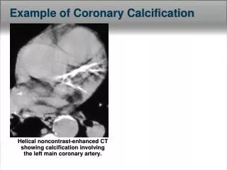
PTE • Filling defect in pulm. art. post-contrast • Widenening, blunting, abrupt termination of arteries • Lung parenchyma distal to thrombus may be oligemic (decrease in vessel number or caliber +/- decrease in pulm. attenuation) • Thrombosis of multiple small pulm aa. – patchy decrease in attenuation (“mosaic oligemia”)
PTE • Pulmonary infarcts – triangular to polyhedral focal region of increased parenchymal attenuation distal to thrombus • “Vascular sign” – thickened or thrombosed vessel leading to apex of infarct • Pleural effusion – often hemorrhagic, likely secondary to pulm. necrosis
“Mosaic oligemia” Pulmonary Infarct
Vascular sign (w/PTE) and infarct PTE and pleural fluid
PTE • False + and false – from insufficient pulmonary artery contrast enhancement • False + from confusing adjacent lymph nodes with art. thrombi
“Pseudothrombus” = right hilar lymphadenopathy (arrowhead), PTE in left descending pulmonary art. (arrow)
“Pseudothrombus” • Streamlining 0 Sec Post 30 Sec Post
NCSU Cases • 10y, MC Silky Terrier • Hepatocarcinoma • Met Check • Normal thorax • link
NCSU Cases • Adjust window and level • Lung parenchyma (W≈1500, L≈-500) • PTE (W≈500, L≈0) • Review transverse, sagittal and dorsal plane images • Review different post-contrast times • Often good pulm. art. opacification at ≈ 1 min. • 1mm images best for small lesions
NCSU Cases • 8 Y, MC, Border Collie • Increased resp. rate • Exercise intolerance • Hypoxemia • Transverse , 75 sec post
NCSU Cases • 10 y, MC Beagle • Suspect glucagonoma • Previous heartworm dz possible • Dorsal
NCSU Cases • 8y, FS, Golden Retriever • Adrenal mass • Hyperadrenocorticism • Dorsal
CT Angiography for Diagnosis of Pulmonary Embolism: State of the ArtSchoepf U, et. al.Radiology, 2004;230:329-337
Surpassed scintigraphy as first-line imaging test for PTE • Many advantages of CT over other imaging • med. and parenchymal struct. can be evaluated • thrombus can be directly visualized • interobserver agreement for CT better than scint. • more cost-effective than algorithms that do not include CT
Historical limitation of this modality - detection of small peripheral emboli (single–detector row CT) • Multislice helical CT= increased speed, decreased motion artifact (entire thorax in single breath hold), increased spatial resolution, isotropic voxels & 3D reformatting • = improved detection of small peripheral emboli
16 slice CT – thrombus in small peripheral vessel w/ associated infarct