Spiral CT in Radiology: Advancements and Challenges
230 likes | 286 Vues
Learn about Spiral CT technology, helical reconstruction, volume scanning, and data acquisition challenges in radiology imaging. Explore the benefits and drawbacks of spiral vs. conventional CT methods.

Spiral CT in Radiology: Advancements and Challenges
E N D
Presentation Transcript
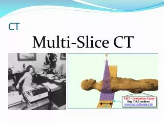
Single Slice Spiral - Helical CT Oh no, not more physics…
Spiral CT • Incentives for development • Shorter study times • Improved 3D imaging • New technology required • Slip ring • Allows continuous gantry rotation
Conventional (Non-spiral) CT • Tube rotates once around patient • Table stationary • data for one slice collected • Table increments one slice thickness • Repeat • Tube rotates opposite direction
Volume Scanning • Data collected continuously • Table moves continuously • Focal spot traces spiral pathwith respect to patient
Helical Reconstruction Complication • Patient moves as gantry rotates • No two fan beams at same z coordinate “z” direction
As Gantry Rotates,Fan Angles Repeat • Distance between repetitions is movement of table during one rotation “z” direction
Data Acquisition Challenges • Projection data not confined to single slice • Streak artifacts • caused by motion • special algorithms required Position at start of rotation Position at start of rotation Position of interest
Calculating Fan Beams at Odd Locationsusing Interpolation • No complete data set for any single z location • Use 2 closest beams in correct orientation • Calculate beam attenuation by interpolating between adjacent beams “z” direction
Spiral Reconstruction Algorithms = real data point • Uses interpolation for • input projection data • output slice attenuation data • Slice can be calculated at any position from raw projection data coordinate of interest Interpolated data
Disadvantage of Interpolation Trust me, interpolation is a guess • Can increase effective slice thickness • Calculation averages data measured at many z values “z” direction
Data Acquisition Challenges • No single slice defined by acquisition geometry • slice localization more difficult • Different slice volume geometry • conventional: cylinder • spiral: wafer with radial crack • Slight increase in effective slice thickness • slice thickness influenced by • fan beam thickness • speed of table motion
Table Moves During Helical Scanning table increment during one rotation Slice Pitch = --------------------------------------- slice thickness Slice thickness TableIncrement
Table Moves During Helical Scanning • Slice thickness determined by collimation • Table motion per revolution determined by table speed table motion during one rotation Slice Pitch = --------------------------------------- slice thickness Slice thickness TableIncrement
Single-Slice Detectors • Many detectors rotate around patient • Single row in z-direction • Slice thickness determined by collimation SliceThickness Z-Axis
Pitch = 1 • Pitch = 1 means patient moves exactly one slice thickness per revolution of tube table motion during one tube rotation Slice Pitch = ---------------------------------------------- slice thickness Beam positions when tube directly above patient
Pitch <1 • Pitch < 1 means patient moves less than a slice thickness during one tube rotation • Can improve visualization of objects table motion during one tube rotation Slice Pitch = ----------------------------------------------- slice thickness Beam positions when tube directly above patient
Pitch >1 • Pitch > 1 means patient moves further than slice thickness during one tube rotation table motion during one tube rotation Slice Pitch = ---------------------------------------------- slice thickness Beam positions when tube directly above patient
Pitch >1 • No gap in image coverage when viewing study table motion during one tube rotation Slice Pitch = ---------------------------------------------- slice thickness No Gap Beam position when tube directly belowpatient
Spiral vs. Conventional CT & Patient Dose • Dose is strongly dependent on pitch Please explain. Inquiring minds wanna know
Pitch = 1 • equivalent dose to non-spiral
Pitch >1 • lower dose for spiral • Table moves faster • Table increment per tube rotation > one slice thickness
Pitch <1 • higher dose for spiral • Table moves slower • table increment per tube rotation < one slice thickness
Pitch & Dose • Dose inversely proportional to pitch • Pitch = 0.5 => Dose doubles • Pitch = 2 => Dose cut in half