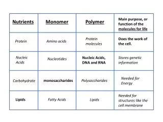
Monosaccharide, Oligosaccharide, and Linkage Analysis
370 likes | 580 Vues
Monosaccharide, Oligosaccharide, and Linkage Analysis. Natasha E. Zachara Ph.D. nzachara@jhmi.edu. What questions might you ask?. What monosaccharides modify my protein? What oligosaccharides modify my protein? How are these sugars linked to each other?

Monosaccharide, Oligosaccharide, and Linkage Analysis
E N D
Presentation Transcript
Monosaccharide, Oligosaccharide, and Linkage Analysis Natasha E. Zachara Ph.D. nzachara@jhmi.edu
What questions might you ask? • What monosaccharides modify my protein? • What oligosaccharides modify my protein? • How are these sugars linked to each other? As always, the choices you make about how you will approach answering these questions will depend in part on the equipment availableto you.
Monosaccharide Analysis • Why? • Knowing which sugars helps to predict the type of oligosaccharides present and thus helps determine the approach to detailed structural analysis • It can also provide the first clues to the presence of a new type of sugar chain. • Step one: release of sugars • Step two: labeling of sugars (if necessary) • Step three: detection and quantification
Sample Preparation • In most cases, salt is an issue: • Proteins can be precipitated to remove salt • Acetone • Methanol • Methanol/Chloroform • Protein can be desalted by Size Exclusion Chromatography • Ammonium formate, ammonium bicarbonate (volatile) • Sephadex-G50, many others • Proteins/peptides purified by reversed phase are also acceptable • Proteins can be separated by SDS-PAGE and blotted to PVDF • Sugars can be desalted • Size exclusion chromatography (time challenging) • Dowex Ion Exchange (in a volatile buffer) • Graphitized Carbon
Release: monosaccharides Acid hydrolysis: • Different sugars and linkages are released at different rates • Low acid: Sialic acid • Neutral and amino sugars: 2M TFA and 4M HCl Harazono A, et al., A comparative study of monosaccharide composition analysis as a carbohydrate test for biopharmaceuticals. Biologicals. 2011 May;39(3):171-80. PMID: 21549615
Detection • The challenge is detecting your released sugars: • PAD: Pulsed Ampometric Detection (no labeling) • Metabolic Labeling • Derivatization for HPLC: • 2-aminopyridine (2AP), • ethyl 4-aminobenzoate (ABEE), • 2-aminobenzoic acid (2AA) • 1-phenyl-3-methyl-5-pyr- azolone (PMP) • 1,2-diamino-4,5-methylenedioxybenzene (DMB) • Capillary Electrophoresis: • 8-aminopyrene-1,3,6-trisulfonate (APTS) • 2-aminoacridone derivatization • Derivatization for GC-MS: • trime- thylsilyl or alditolacetate Harazono A, et al., A comparative study of monosaccharide composition analysis as a carbohydrate test for biopharmaceuticals. Biologicals. 2011 May;39(3):171-80. PMID: 21549615
Separation • HPLC • Anion Exchange Chromatography (HPAEC) • Reversed Phase • Ion Exchange (TSK Fractogel) • Capillary Electrophoresis • GC-MS
High Performance Anion Exchange Chromatography-Pulsed Ampometric Detection (HPAEC-PAD) • Advantages: • No Labeling • No interference from labeling reagent • Reasonable sensitivity (10pmol) • Disadvantages: • Specialized HPLC (All hydroxide) • The Separation: • Under alkaline conditions, the hydroxyl groups are ionized to oxyanions • A monosaccharide possesses several ionizablehydroxyl groups with the following hierarchy of acidity: 1-OH > 2-OH >> 6-OH > 3-OH > 4-OH • The varying location of the OH groups results in slight differences in the pKa value (ranging from 12 to 14) of individual monosaccharides Harazono A, et al., A comparative study of monosaccharide composition analysis as a carbohydrate test for biopharmaceuticals. Biologicals. 2011 May;39(3):171-80. PMID: 21549615
HPAEC-PAD Columns • CarboPAcPA1, 10, 20 • Monosaccharides • Disaccharides • Sialic acid • CarboPAc PA 100, 200 • Oligosaccharides • CarboPAcMA1 • Aliditols (monosaccahrides, oligosaccharides) Behan JL, Smith KD. The analysis of glycosylation: a continued need for high pH anion exchange chromatography. Biomed Chromatogr. 2011 Jan;25(1-2):39-46. PMID: 20821735
How does pad work? Ampometric Detection: • Any analyte that can be oxidized or reduced is a candidate for amperometricdetection • A voltage (potential) is applied between two electrodes positioned in the column effluent • The measured current changes as an electroactiveanalyte is oxidized at the anode. Pulsed Ampometric Detection: • Important as it ensures that the gold electrode does not become coated by oxides • In pulsed amperometric detection (PAD), a working potential is applied for a short time (usually a few hundred milliseconds), followed by higher or lower potentials that are used for cleaning the electrode • The detection of monosaccharides and amine-containing glycoconjugates with low levels of glycosylation can be compromised from fouling of the working electrode by amino acids.
Examples: monosaccharides http://www.dionex.com/
Examples: monosaccharides Note, that Sodium Acetate is required to elute sugar phosphates http://www.dionex.com/
Sugar Alditols Note, that a gradient of sodium hydroxide is required http://www.dionex.com/
HPAEC: SIALIC acid The strong charge requires more robust conditions; an increasing sodium acetate linear gradient is useful (50–250 mm over 30 min . O-Acetyl groups can be labile, andcansaponify during analysis. There are alternative non-high pH based methods. http://www.dionex.com/
HPAEC: Neutral oligosaccharides • Separation of oligosaccharide is based on size, charge, monosaccharide composition and intra-chain linkages • Separation is based on a sodium acetate gradient; the greater the negative charge (greater the number of NeuAc) and number of sugars, the higher the sodium acetate concentration required http://www.dionex.com/
HPAEC: Charged Oligosaccharides the greater the overall negative charge, the greater the retention time. http://www.dionex.com/
Desalting post-HPAEC Packer NH, Lawson MA, Jardine DR, Redmond JW. Glycoconj J. 1998 Aug;15(8):737-47. A general approach to desalting oligosaccharides released from glycoproteins. PMID: 9870349
GC-MS: monosaccharides • Sugars must be derivatized: trimethyl esters, aldlitol acetates, permethylation • Also possible to release sugars by methanolysis, and derivatize to TMS derivatives • Detection: Flame Ionization detector • Several types of stationary phases can be used for the analysis of acetylated alditols: • CP-Sil 5 WCOT, SP-1000, Silar 10C, OV-1, SE-54, 5% DB-5, DB-1, Carbowax 20M, SE-30, OV-101, and OV-275. • Other stationary phases used for the analysis of trimethylsilylated glycosides and methyl glycoside methyl esters include CP-Sil 5 WCOT, 5% DB-5, DB-1, SE-30, and OV-101. PMID: 6057635
GC-MS: monosaccharides • Identify sugars by retention time and fragmentation patterns • Lose some information re: modifications of sugars • Quantitative PMID: 6057635
HPLC: monosaccharides Labeling with different derivatives: A: 2AP (2-aminopyridine ) B: ABEE (ethyl 4-aminobenzoate ) C: 2AA (2-aminobenzoic acid ) D: PMP (1-phenyl-3-methyl-5-pyrazolone ) Separation Technologies: Strong Anion Exchange (TSK Fractogel) Reversed Phase (C18) CE Great sensitivity… Derivatization can fluctuate in efficiency May be necessary to include a clean up step Longer run times Harazono A, et al., A comparative study of monosaccharide composition analysis as a carbohydrate test for biopharmaceuticals. Biologicals. 2011 May;39(3):171-80. PMID: 21549615
Capillary Electrophoresis (CE) Separation buffers: 50 mM sodium phosphate (pH 5.5) or 150 mM sodium borate-50 mM sodium phosphate (pH 7.0) running buffer. Run times are fast Compatible with mass spectrometry Not preperative Abo M, He LP, Sato K, Okubo A. Determination of monosaccharidesderivatized with 2-aminobenzoic Acid by capillary electrophoresis. Methods Mol Biol. 2013;984:45-50. doi: 10.1007/978-1-62703-296-4_4. PMID:23386335
Oligosaccharides • There are three steps: • Step one: Release • Step two: Detections • Step three: Separation
Release: oligosaccharides • Reductiveb-elimination: • releases O-linked sugars, but the carbohydrates must be reduced to prevent peeling. • results in sugar alditols, which are not compatible with many labeling techniques • Alditol acetates are volatile and are easily detected by GC-MS. • Hydrazinolysis: • Hydrazine hydrolysis is an effective method for the complete release of unreduced O- and N-linked oligosaccharides. • not as popular as other techniques as it’s toxic and can affect the integrity of the peptide/protein backbone. • Enzyme hydrolysis: • PNGaseF releases most N-linked glycans.
Detection: Oligosaccharides • Radiolabels: • Metabolic Labeling • Glycosyltransferases • b-elimination • Reductive amination: • Incorporation of fluoresecent derivatives (see above) • Pulsed amometric detection • Mass spectrometry (GC, MALDI, ESI-MS)
Separation • Thin Layer Chromatography • High-voltage Borate Paper electrophoresis • FACE • Size exclusion/Gel Permeation Chromatography: • HPLC • Graphitized Carbon • Reversed Phase • HILIC • HPAEC • GC-MS • Mass Spectrometry • Can be combined with HPLC
HPLC: Graphitized Carbon • Not entirely clear how graphitized carbon separates oligosaccharides, although size, shape and charge are all important • Can separate anomers • N-linked sugars should be reduced to avoid peak splitting • Sensitive to the low femtomolelevel • Columns require significant regeneration Karlsson NG, Wilson NL, Wirth HJ, Dawes P, Joshi H, Packer NH. Rapid Commun Mass Spectrom. 2004;18(19):2282-92. Negative ion graphitised carbon nano-liquid chromatography/mass spectrometry increases sensitivity for glycoprotein oligosaccharide analysis.
HPLC: hydrophilic interaction liquid chromatography (HILIC) • HILIC: Sugars are separated by a partitioning mechanism in contrast to the traditional adsorption chromatography on normal phase materials • Typical equilibration conditions 10–25% water in acetonitrile with a low concentration of acid or salt (mostly below 100 mM) • Common columns include: TSK Gel-Amide 80 and ZIC-HILIC-SPE • Compatible with spectrometry • Detection requires derivatization
Biogel p4 • Carbohydrates can be separated by size • Typically Biogel P-2, P-4 and P-6 (<400 mesh resin) are used. • BiogelP-4 is best for the separation of 3-24 units • Sugars are detected by the incorporation of a radiolabel or a fluorescent tag • For O-linked/N-linked sugars it is possible to introduce this tag during reductive b-elimination, by using tritiated sodium borohydride • The carbohydrates can also be detected using colorimetric assays Columns are calibrated and retention time expressed in units of glucose.
FACE: fluorophore-assisted carbohydrate electrophoresis • Separate oligosaccharides by electrophoresis • Addition of a tag (by reductive amination) provides a charge and a meads of detection • Separation may vary if sugars are sulfated, acetylated of sialylated as this will change the size: charge ratio. • Can be combined with enzyme treatments to look at the structure of the oligosaccharides, however, this requires purified oligo’s
GC-MS: Oligosaccharides • Large oligosaccharides (>10) are difficult to detect • Must use alternative approaches, eg. FAB-MS • Typically permethylated Karlsson N G et al. J. Biol. Chem. 1997;272:27025-27034
Fragmentation: Oligosaccharides Karlsson N G et al. J. Biol. Chem. 1997;272:27025-27034
Linkage Analysis • What questions are you really asking? • Which sugars are linked to each other, and in what order • Though which hydroxyl residues are these sugars linked • What is the anomericity of the linkage? • Methods: • NMR (best, but requires largish amounts of pure material) • Exoglycosidase digestion of oligosaccharides (requires the right enzymes) • Methylation Analysis: • GC-MS • ESI-MS (best with upstream separation of oligosaccharides) • You may get some information about anomericity when this is performed in tandem with column chromatography • MALDI-TOF • Mass Spectrometry
Methylation Analysis Methylation analysis is a well-established method for determining linkage positions. Introduce a stable substituent (an ether-linked methyl group) onto each free hydroxyl group of the native glycan Acid cleavage (glycosidic linkages and more labile than ether-linked methyl groups) Partially methylated monosaccharides with free hydroxyl groups are derivatized to produce volatile molecules amenable to GLC-MS analysis. Typically, monosaccharides are reduced (no alpha or beta anomers) followed by acetylation of the free hydroxyls Detection by GC-MS or ESI-MS No sequence information, no anomeric linkage information Essentials of Glycobiology Second Edition Chapter 47, Figure 3
Methylation Also Improves Assignment DuringMass Spectrometry What Mass Spectrometry can do: 1) heterogeneity and type of glycosylation; 2) sites of glycosylation; 3) glycan-branching patterns; 4) the number and lengths of antennae, their building blocks, and the patterns of substitution with fucose, sialic acids, or other capping groups such as sulfate, phosphate, or acetyl esters; 5) complete sequences of individual glycans. What it can’t do: Which Glucose versus Mannose Anomericity How does Methylation Help: Better data Greater confidence in linkage analysis by MS
Wednesday • This will be a demonstration laboratory: • You will set up one protein for sequencing (and analyze the cycles) • We will talk about strategies for site-mapping proteins (mini-lecture) • You will analyze some spectra of O-GlcNAc modified peptides
Reading • Harazono A, et al., A comparative study of monosaccharide composition analysis as a carbohydrate test for biopharmaceuticals. Biologicals. 2011 May;39(3):171-80. PMID: 21549615 • Behan JL, Smith KD. The analysis of glycosylation: a continued need for high pH anion exchange chromatography. Biomed Chromatogr. 2011 Jan;25(1-2):39-46. PMID: 20821735 • Manzi AE. 2001 May;Chapter 17:Unit17.19A. Total compositional analysis by high-performance liquid chromatography or gas-liquid chromatography. PMID: 18265151 • Manzi AE, Diaz S, Varki A. Anal Biochem. 1990 Jul;188(1):20-32. High-pressure liquid chromatography of sialic acids on a pellicular resin anion-exchange column with pulsed amperometric detection: a comparison with six other systems. PMID: 2221361 • Reinhold V, Zhang H, Hanneman A, Ashline D. Mol Cell Proteomics.Toward a platform for comprehensive glycan sequencing. 2013 Apr;12(4):866-73. PMID: 23438731 • Hansson GC, Karlsson H. Methods Mol Biol. Gas chromatography and gas chromatography-mass spectrometry of glycoprotein oligosaccharides. 1993;14:47-54. PMID: 8348243 • Kim JH, Shome B, Liao TH, Pierce JG. Anal Biochem. Analysis of neutral sugars by gas-liquid chromatography of alditol acetates: application to thyrotropic hormone and other glycoproteins. 1967 Aug;20(2):258-74. PMID: 6057635