EXTREMITY TRAUMA
EXTREMITY TRAUMA. Instructor Name: Title: Unit:. OVERVIEW. Relationship of extremity trauma to assessment of life-threatening injury Types of extremity injuries Assessment & management General Estimation of blood loss Splinting Specific injuries. FRACTURE PRIORITIES.

EXTREMITY TRAUMA
E N D
Presentation Transcript
EXTREMITY TRAUMA • Instructor Name: • Title: • Unit:
OVERVIEW • Relationship of extremity trauma to assessment of life-threatening injury • Types of extremity injuries • Assessment & management • General • Estimation of blood loss • Splinting • Specific injuries
FRACTURE PRIORITIES • Fractures rarely life-threatening • Perform BTLS Primary Survey to find life-threatening injuries • Do not be distracted by obvious but not life-threatening extremity injuries • Be alert to major bleeding from extremity injuries
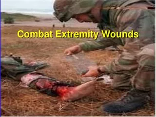
TYPES OF FRACTURES • Open • Bone ends protrude through the wound • High risk of infection • Closed • No opening through the skin • Fractures may • Damage adjacent nerves and vessels • Produce severe bleeding • Blood loss may be internal
DISLOCATIONS • Joint deformity may be fracture or dislocation • Can cause neurovascular compromise of distal extremity • Always assess • Distal sensation • Distal motor function • Distal pulses and skin color
AMPUTATIONS • Control bleeding by direct pressure • Tourniquets rarely needed • Locate amputated part • Do not place amputated part directly in ice or water • Place part in plastic bag • Place bag in ice-water mixture
SPRAINS & STRAINS • Signs similar to fractures • X-rays needed to distinguish from fractures • Treat as if fractured “If an extremity hurts, immobilize it”
OPEN WOUNDS • Control bleeding with pressure • Tourniquets rarely needed • Check distal PMS • Pulse • Motor • Sensory COURTESY ROY ALSON M.D.
IMPALED OBJECTS • Stabilize in position found • Removal may cause uncontrollable bleeding • Exceptions • Object in cheek • Cannot control major bleeding with object in place
Early Pain Paresthesias Late Pallor Pulselessness Paralysis COMPARTMENT SYNDROME Pathophysiology Signs and symptoms
SIGNS & SYMPTOMS OF EXTREMITY INJURY • Pain • Deformity • Swelling • Loss of movement • Crepitus COURTESY ROY ALSON, M.D.
ASSESSMENT • Scene Size-Up • Clues to specific injuries • BTLS Primary Survey • Pelvic fractures or bilateral femur fractures are Load & Go • Control major bleeding • History may suggest other injuries
BLOOD LOSS FROM FRACTURES • Pelvis - 500cc for each break • May lacerate major vessels causing major internal bleeding • Femur - 1000cc • Multiple fractures can produce life-threatening hemorrhage • May all be internal
Deformities Contusions Abrasions Penetrations Burns Tenderness Lacerations Swelling DETAILED EXAMCHECK EXTREMITIES FOR ALSO CHECK FOR PMS
MANAGEMENT • SPLINTING • Decreases pain • Prevents further injury • Decreases blood loss COURTESY DAVID EFFRON, M.D.
GENERAL RULES OF SPLINTING • Visualize injured part • Check and record PMS before and after splinting • May apply gentle in-line traction • Cover open wounds with sterile dressings • Pad the splint • Immobilize one joint above and below the site of the injury
GENERAL RULES OF SPLINTING • Do not push bone ends back under the skin • May apply splints en route to the hospital • If in doubt, splint • Never delay transport of critical patient to perform splinting of minor fractures
MANAGEMENTLOAD & GO PATIENTS • Spinal immobilization • Long backboard • C-collar • Head immobilizer • Limit splinting until en route • Backboard acts as “whole body” splint
MANAGEMENTSPECIFIC INJURIES • CLAVICLE FRACTURES • Common injury • Apply sling & swathe
SHOULDER INJURIES • AC separation • Sling & swathe • Shoulder dislocation • Use pillow with sling & swathe • Fracture • Use sling & swathe
ELBOW INJURY • Fracture or dislocation may cause neurovascular injury • Splint in position found • Transport promptly
FOREARM/WRIST INJURY • Rigid splint • Keep hand in position of function • Air splint • May be difficult to reassess circulation • Pillow
FEMUR FRACTURES • High force injury • High potential for shock • May use traction splint • PASG or air splint may give adequate stabilization COURTESY OF ROY ALSON M.D.
KNEE FRACTUREOR DISLOCATION • Orthopedic emergency • Frequently causes vascular injury • Dislocation associated with high incidence of leg amputation
MANAGEMENT KNEE DISLOCATION • Obvious dislocation without distal pulse • Apply gentle in-line traction • If gentle traction does not restore the pulse • Splint in place • Prompt transport
Frequently open fractures Significant hemorrhage possible Dress open wounds Depending on level of fracture Upper - Rigid splint Lower - Air splint or pillow TIBIA-FIBULA FRACTURES COURTESY OF ROY ALSON M.D.
FOOT OR HANDINJURIES • Common industrial injury • Often disabling • Rarely life-threatening • Splint foot with pillow • Splint hand in position of function
SUMMARY • Note mechanism of injury • Remember priorities • ABCs first • Be prepared for shock • Record PMS
SUMMARY • Critical patients • Do not waste time on minor splinting • Immobilize spine • Apply other splints en route • Immobilize one joint above and below • If in doubt, splint