Peritoneum: An Overview for Medical Professionals
Explore the general features of the peritoneum in the abdominal and pelvic cavities, its layers, and the role it plays in organ protection and movement. Learn about intraperitoneal and retroperitoneal organs and the divisions of the peritoneal cavity. Discover the functions of the greater omentum, lesser sac, and peritoneal ligaments, omenta, and mesenteries. Gain insights into the anatomy and attachment points of the falciform ligament and its significance in liver connectivity.

Peritoneum: An Overview for Medical Professionals
E N D
Presentation Transcript
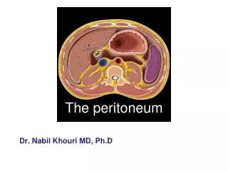
Dr. NabilKhouri MD, Ph.D The peritoneum
The peritoneum • General features • The peritoneum is a thin serous membrane that line the walls of the abdominal and pelvic cavity and cover the organs within these cavities • Parietal peritoneum-lines the walls of the abdominal and pelvic cavities • Visceral peritoneum -covers the organs • Peritoneal cavity -IS the potential space between the parietal and visceral layer of peritoneum, in the male, is a closed sac, but in the female, there is a communication with the exterior through the uterine tubes, the uterus, and the vagina • This potential space between the two layers is filled with approximately 100 mL of very thin film of serous fluid to facilitate the movement of the abdominal organs.
Intraperitoneal organ means that the organ is completely covered by visceral layer of peritoneum Retroperitonealorgan means that the organ lies behind the peritoneum and partially covered by visceral peritoneum e.g. pancreas, ascending & descending colon.
The peritoneal cavity Is divided into two main sacs: 1- Greater sac. 2- Lesser sac or OMENTAL BURSA. These two sacs are interconnected by a single oval opening called the EPIPLOIC FORAMEN or opening into lesser sac or foramen of Winslow The Greater sac It is the part of peritoneal cavity which lies behind the anterior abdominal wall. Peritoneum lines the Anterior abdominal wall then the under surface of diaphragm, from where it is reflected on to superior surface of liver forming the upper layer of coronary ligament of the liver
The Lesser sac A peritoneal pouch lies behind stomach & lesser omentum It projects upwards as far as the diaphragm “superior recess”. Inferiorly it lies within the folding of the greater omentum. Its lower part is usually obliterated due to reflection of the anterior & post layers of the greater omentum.
Upper border: Extends from porta hepatis, fissure for ligamentum venosum to lower end of esophagus. • Lower border: Inferior margin of greater omentum. • Lt border: Lt margin of greater omentum, gastrosplenic & lienorenal ligaments. • Rt border: Rt. Margin of greater omentum, opening into lesser sac.
Greater omentum Four-layered fold of peritoneum, the anterior two layers descend from the greater curvature of stomach and superior part of duodenum and hangs down like an apron in front of coils of Small intestine, and then turns Upward and attaches to the transverse colon. If an infection occurs in the intestine, plasma cells formed In the lymph nodes combat the infection and help prevent it from spreading to the pritoneum. It has a role in immunity and is sometimes referred to as the ‘abdominal policeman’ because it can migrate toinfected viscera.
The Lesser Omentum Connects liver to stomach attached above to porta hepatis & fissure for ligamentum venosum inferiorly to lesser curvature of the stomach,& 1st inch of duodenum. Its free margin contains: Portal vein: Posterior Bile duct : Anterior & right Hapatic artery: anterior &left Gastrosplenic ligament Connects the stomach to the hilum of the spleen Splenicorenal or lienorenal ligament Connects the hilum of spleen to front of the left kidney.
Peritoneal ligaments Two layers of peritoneum that connect viscera to abdominal walls. Falciform, coronary, right & left triangular ligaments Omenta Two layers of peritoneum that connect stomach to another viscus. Lesser & greater omenta and gastrosplenicomentum (ligament). Mesenteries Two layers of peritoneum connecting small intestine to post abdominal wall. It has 2 borders 1- Attached border: to post abdominal wall & 2- Free border: which encloses the jejunum & ileum. Vessels, nerves. Lymphatic enter small intestine between the two layers.
The Falciform Ligament A sickle-shaped fold of peritoneum connects the Anterior Abdominal Wall with the liver slightly to the right of the median plane. Ant border: Attached to under surface of diaphragm & anterior abdominal wall Post border: Attached to sup & ant surfaces of liver Free margin connects the umbilicus to liver it contains the ROUND LIGAMENT of the liver or (Ligamentumteres).
Then, it descends from sup surface of liver to the ant. surface then inferior surface of liver. From the post. part of inferior surface peritoneum reflected on to front of right kidney & RT suprarenal gland forming the lower layer of coronary ligament. The lower & upper layers of coronary ligament bound a large area on the post surface of the liver called BARE AREAof the liver which has no peritoneal covering. Upper part of coronary ligament Lower part of coronary ligament
Peritoneal Recesses, Spaces,and Gutters Duodenal Recesses there may be four small pocket like pouches of peritoneum called the superior duodenal, inferiorduodenal, paraduodenal,and retroduodenal recesses. Cecal Recesses three peritoneal recesses called the superior ileocecal, the inferior ileocecal, and the retrocecal recesses.
Intersigmoid Recess The intersigmoid recess is situated at the apex of the inverted V-shaped root of the sigmoid mesocolonits mouth opens downward. • Subphrenic Spaces The right and left anterior subphrenicspaces lie between the diaphragm and the liver, on each side of the falciform Ligament. The right posterior subphrenic space lies between the right lobe of the liver, the right kidney, and the right colic flexure. The right extraperitoneal space lies between the layers of the coronary ligament and is therefore situated between the liver and the diaphragm. • The paracolic gutters lies lateral and medial to the ascending and descending colon respectively.
Innervation of peritoneum: Parietal peritoneum is sensitive to pain, pressure, temperature & touch,(pptt) Parietal peritoneum is supplied by: Lower 6 thoracic nerves (T7-- T12) First lumber nerve ( L1) Central part of diaphragmatic parietal peritoneum is supplied by phrenic nerve. Visceral peritoneum is sensitive to stretch & tearing. It is supplied by autonomic afferent nerves which supply the viscera. NB. Parietal peritoneum of the pelvis is supplied by Obturator nerve.