Peritoneum
This article explores the peritoneum's anatomy, its ontogenesis, and the role of the coelom during the second month of embryonic development. Key structures such as the ventral and dorsal mesenteries, major and minor omenta, as well as the various layers and types of organs within the abdominal cavity—intraperitoneal, mesoperitoneal, and extraperitoneal—are discussed. Additionally, the function and structural organization of the peritoneal cavity are analyzed, including important ligaments and the relationship between internal organs. Clinical implications like hysterosalpingography and common malformations are also addressed.

Peritoneum
E N D
Presentation Transcript
Ontogenesis coelom • 2-nd month. The source - splanchnopleura • Ventral mesentery • Dorsal mesentery • Major and minor omentums
Abdominal cavity Walls • Fascias • Spaces
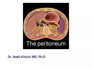
Peritoneum Parietal layer Visceral layer
Abdominal cavity- • this space of the body, located lower than the diaphragm is filled with internal organs: predominantly digestive and urogenital systems
Walls of the abdominal cavity Walls of the peritoneal cavity • Superior-diaphragma • Anterolateral –abdominal muscles • Posterior – vertebral column and posterior abdominal wall • Inferior – perineum
Functions of the peritoneum • Fixation of internal organs • ligaments,mesentery, omentum • Motions of internal organs • Isolation of internal organs • Reflex area • Trophic • Protection • Regeneration • Reconstructive surgery
Hysterosalpingogram • Radiograph of the uterus and uterine tubesafter injection of radiocontrast material into the uterus through uterine ostium
LIGAMENTS PRIMARY SECONDARY
Division of the peritoneal cavity • Upper storey • Middle storey • Lower storey
Upper storey • above - diaphragm; • anteriorly - parietal peritoneum of anterior abdominal wall; • below – colon transversum and its mesentery • posteriorly - posterior abdominal wall;
Upper storey • Bursa hepatica • Bursa pregastrica • Bursa • omentalis • - Foramen epiploicum
Bursa hepatica • Above – diaphragm • Left side – lig. falciforme • Anteriorly - parietal peritoneum of anterior wall • Behind - right kidney, adrenal gland
BURSA PREGASTRICA • Above – diaphragm • Anteriorly- parietal peritoneum of anterior wall • Right side - lig. falciforme • Behind – stomach • Left side – spleen, its ligaments
BURSA OMENTALIS • Above - caudate lobe of the liver • Anteriorly – lesser omentum, the posterior wall of the stomach • Behind- parietal peritoneum • Below - mesentery of the transverse colon • Left side -ligaments of the spleen
FORAMEN EPIPLOICUM • Above - caudate lobe of the liver • Anteriorly - lig. hepatoduodenalis • Below – pars superior duodeni • Behind- parietal peritoneum
Middle storey • Mesocolon • Linea bispinata
Mesenteric sinuses • RIGHT • LEFT
Lateral canals • RIGHT • LEFT
Recesses • recessus duodenalis superior and inferior (1) • recessus ileocecalissuperior and inferior (2,3) • recessus retrocecalis(4) • recessus intersigmoideus (5) 1 2 3 4 5
Lowerstorey • Excavation • Rectovesicalis • Rectouterina • Vesicouterina
Retroperitoneal spaces • Paracolon • Paranephron
Malformations REMNANT OF VENTRAL MESENTERIUM DEFECT OF GREATER OMENTUM
Malformations RUDIMENTAL OMENTAL FIMBRIA OF GREATER CURVATURA ATRESIA OF TRANSVERSE COLON