Peritoneum
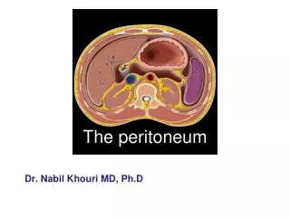
Peritoneum. Peritoneum It is divided into two parts : 1 – Visceral : Surrounding the viscera. 2 – Parietal : lining the rest of the cavity.

Peritoneum
E N D
Presentation Transcript
Peritoneum It is divided into two parts : 1 – Visceral : Surrounding the viscera. 2 – Parietal : lining the rest of the cavity. The parietal portion is richly suppliedwith nerves and when irritated, produce severe pain accurately localized to the affected area. The visceral peritoneum on the other hand is poorly supplied with nerves and pain arising there from is vague and badly localized.
Surgical physiology : The peritoneal cavity is the largest cavity in the body, the surface area of its lining membrane being nearly equal to that of the skin. It is composed of two layers, first one is flattened polyhydral cells which is one layer thick and the other one is fibroelastic tissue. Bothe these layers form the peritoneum. Beneath the peritoneum lies a network of lymphatics and rich plexuses of capillaries from which all absorption and exudation occur. In normal condition, only few cc of pale yellow fluid is present in peritoneal cavity for lubrication of mobile viscera.
Acute peritonitis : Nearly all verities of peritonitis are due to an invasion of the peritoneal cavity by bacteria. Most cases of bacterial peritonitis are polymicrobial, and usually both aerobic and anaerobic organisms are present. The exception is primary peritonitis (spontaneous peritonitis) in which a pure infection with streptococcal, pneumococcal or haemophilus organisms is seen. Bacteriology : 1 – Bacteria from the alimentary tract : usually the infection is caused by two or more strains. The commonest invaders are E.Coli, Anaerobic strept, and bacteroides. Less frequently Clostridium Welchii, Staph or Klebsiella Pneumoniae. 2 – bacteria not from alimentary tract : Peritonitis due to Gonococcus, Beta-haemolytic strept, Pneumococcus and Mycobacterium Tuberculosis.
Baths of bacterial invasion of peritoneal spaces: 1 – Direct infection : - Via perforation of some parts of GIT . - Through a penetrating wound of the abdominal wall. - Operative e.g. drains, foreign materials ….etc. 2 – local extension : - From an inflammed organ e.g. appendicitis. - Migration through gut wall e.g strangulated hernia. - From or via fallopian tubes. 3 – Blood – stream : - part of general septicemia. * Even an initially sterile peritonitis ( e.g due to rupture gall bladder ) soon it becomes infected by transmigration of organism from the bowel which can takes a few hours only.
Natural factors that favour localization of peritonitis : 1 – Anatomical : The greater sac of peritoneum is divided into the pelvic and the peritoneal cavity proper. The later is redivided into a supracolic and an infrcolic compartments by the transverse colon and transverse mesocolon, which prevent the spread of infection from one to other. Also other adhesions will affect the spread of infection like Rt and Lt paracoloic gutters. 2 – Pathological : a/ Flakes of fibrin appear and causes coils of intestine to become adherent to one another and to parietes. b/ Outpouring of serous fluid rich in leucocytes and antibodies that soon become turbid. c/ Peristalisis is reduced in affected coils to prevent distribution of infection. d/ Greater omentum will try to envelop and become adherent to inflamed structure and will form a barrier for spread of infection.
3 –Surgical : Drains are frequently used postoperatively to assist localization of intra-abdominal collections Natural factors that tend to cause diffusion of peritonitis: 1 – A prime factor in the spread of peritonitis is weather it develops rapidly or slowly : -A sudden perforation of inflamed appendix or other hallow viscus before protective mechanisms. - Perforation proximal to an obstruction. 2 – The ingestion of food, or even water will stimulate peristalsis and will hinders localization. 3 – Severity of virulence of causative organism. 4 – In children the omentum is small. 5 – Rough handling of localized collection by surgery can cause spread . 6 – Deficient natural resistance ( immune deficiency )
Clinical features : Localized peritonitis : 1 – To start with it is bound up immediately to the causative lesion, and the initial symptoms and signs are those of that lesion. 2 – Elevated temperature and increased pulse rate. 3 – Severe abdominal pain. 4 – Vomiting. 5 – Guarding and rigidity ( the most important signs ) over the affected area only with a positive release sign. 6 – Shoulder tip pain in cases of subphrinic peritonitis. 7 – Tenderness on PR or PV in pelvic peritonitis.
Generalized peritonitis : Initial phase : sever pain made worse by movement or breathing which is first experienced at the site of original lesion and then spread outward. Vomiting, tenderness and rigidity which are generalized. Increased pulse and temperature. Intermediate phase : Peritonitis may resolve so that pulse slows, pain and tenderness diminishes, leaving a silent soft abdomen. These are features that can easily mislead the observer. The condition may localize producing one or more abscesses with overlying swelling and tenderness. Terminal phase : if resolution or localization not happen, the abdomen remains silent and increasingly distends. Circulatory failure will happen with cold extremities, sunken eyes, dry tongue, thready pulse, drawn and anxious face ( Hippocratic face ) then patient become unconscious.
Investigations : Usually clinical examination is more important for making diagnosis of peritonitis, but we have some tests to be done : 1 – Leucocytosis. 2 – Peritoneal aspiration : which can give us an indication about the cause such as perforated DU with bile aspiration, bleeding with if the aspirate is blood …ect. 3 – Radiography of abdomen : may reveal free air or confirm the presence of dilated bowel loops with multiple air fluid levels. 4 – Serum amylase to exclude pancreatitis. 5 – Ultrasound to exclude presence of collection of fluis, pus or blood.
Treatment : It consist of three lines of treatment : 1 – General care of the patient. 2 – Neuralization of local sources. 3 – Peritoneal lavage. 1 – General care of the patient : a/ Intravenous fluid : Patient is hypovolemic and have electrolyte disturbence. Central venous line is important for monitoring. Also plasma protein depletion need correction. If recovery of patient delayed for 7-10 days, intravenous feeding ( Paranteral nutrition ) is mandatory. b/ A nasogastric tube : intermittent aspiration till paralytic ileus resulted from peritonitis is resolved.
c/ Antibiotics : Adminstration of antibiotic to prevent the multiplication of bacteria. As the infection is mixed one so antibiotic against aerobic and anaerobic micro-organisms should be used. d/ A fluid balance chart : Input and output should be calculated so the intake and loss be equal. Haematocrit and serum electrolyte and blood urea must be checked. e/ Analgesia : Patient should be nursed in semi sitting position and pain killer should be given. Freedom from pain allow early mobilization and early physiotherapy to prevent deep venous thrombosis and pulmonary embolism.
2 – Neutralization of local source : this means dealing with the cause of peritonitis because treating the cause will prevent increase severity of the condition. As appendicectomy, or dealing with perforated DU …ect. 3 – Peritoneal lavage : After dealing with the cause we should do peritoneal lavage to remove the toxic material from the peritoneal cavity for early recovery.
Prognosis : With modern treatment diffuse peritonitis carries a mortality of about 10%. The lethal factors are : 1 – Bacterial toxaemia. 2 – Paralytic ileus. 3 – Bronchopneumonia. 4 – Electrolyte imbalance. 5 – Renal failure. 6 – Undrained collection. 7 – Bone – marrow suppression. 8 – Multisystem breakdown. Post – operative peritonitis has a mortality of at least 50%. Faecal peritonitis has a mortality rate of about 75%. Peritonitis associated with appendicitis, perforated peptic ulcer or bile leakage are much less dangerous and carry a mortality of less than 10%.
Complications of peritonitis : 1 –Acute intestinal obstruction due to peritoneal adhesions : This gives central abdominal colicky pain with radiological evidence of gas and fluid levels confined to the upper portion of the small intestine. 2 –Parylitic ileus : There is usually little pain and gas lops with fluid levels are seen distributed throughout the small and large intestine on the radiography. 3 –Residual abscesses : Abscess formation following local or diffuse peritonitis. The symptoms and signs of a purulent collection may be very vague and consist of lassitude, anorexia, pyrexia ( low grade ), tachycardia, leukocytosis and localized tenderness with or without palpable mass.
These abscess can be treated conservatively to start with and by close observation of the patient till it will be adherent to abdominal wall that its drainage dose not need opening of peritoneal cavity or if patient condition deteriorate, urgent drainage should be done. Some deeper abscesses can be drained by placement of drainage tube under fluoroscopic or ultrasonographic control. The other and the last option is surgical drinage by operning the peritoneal cavity.
Pelvic abscess It is the most common site of an intraperitoneal abscess. Because the appendix is often pelvic and the fallopian tubes are frequent loci of infection. The most characteristic features of this abscess is the diarrhea and passage of mucous in stool. Rectal examination reveal a bulging of the anterior rectal wall. This abscess can rupture through the rectum leading to resolution of the condition or it should be drained per rectally or per vaginally in female through the posterior fornix.
Subphrenic abscesses Anatomically there is four intraperitoneal and three extrperitonealsubphrenic spaces. Three of these spaces are on either side of the body and one approximately in the midline. I Left superior intraperitoneal space ( left subphrenic ): Bounded by : - Above : diaphragm. - Behind : left triangular ligament and left lobe of liver, gastrohepaticomentum and anterior surface of stomach. - Right : falciform ligament. - Left : spleen, gastrosplenicomentum and diaphragm. * The commonest causes of abscesses here are operations on stomach, tail of pancreas, spleen or splenic flexure of colon.
II Left anterior I ntraperitoneal space ( left subphrenic ) : Also called the lesser sac. The commonest type of suppuration here is the pancreatic pseudocyst. III Right superior intraperitoneal ( Right subphrenic ) : It is between the right lobe of the liver and the diaphragm. Bounded by : - Anteriorly : anterior layer of coronary ligament and right triangular ligament. - left : falciform ligament. The commonest cause of abscess here is perforating cholecystitis, perforated duodenal ulcer and appendicitis.
IV Right inferior intraperitoneal ( Right subphrenic ) : Beneath the right lobe of liver in Rutherford Morison’s pouch. Bounded by : - Right : right lobe of liver and the diaphragm. - Left : Foramen of winslow. - In front : liver and gall bladder. - Behind : The upper part of right kidney and the diaphragm. It is the deepest space of all the four and the most common site for subphrenic abscess. It is usually due to appendicitis, cholecystitis and perforated duodenal ulcer.
V Right and left extraperitoneal spaces These spaces are the right and left perinephric abscesses. VI Midline extraperitoneal which is the bare area of the liver which can develop abscess in amoebic hepatitis or pyogenic liver abscess. Clinical Features : A patient with intra-abdominal problem ( ex perforated DU ) and the condition of patient not improve after that with constitutional symptoms of tiredness, pyrexia, loss of appetite…etc. and abdominal examination may reveal tenderness, rigidity or grading or even palpable swelling.
Investigations : 1 – Blood count : leucocytosis. 2 – Plain radiography : may demonstrate air under diaphragm. 3 – U/S or CT – Scan. Treatment : 1 – aspiration or insertion of drain under ultrasound guide but caution should be taken for not to insert needle through the chest that will cause spread of infection to the pleural space. 2 – Surgical drainage over the maximum tenderness and welling which is better to be done by extraperitoneal approach.