Download

1 / 75
760 likes | 958 Vues
Forelimb Imaging Quiz. Developed by: Sorcha McCaughley & Mark Brims Approved by: Alison King & Maureen Bain Supported by: The Chancellor’s Fund. Forelimb Imaging Quiz. START! Developed by: Sorcha McCaughley & Mark Brims Supported by: The Chancellor’s Fund. Set the scene….

E N D
Forelimb Imaging Quiz Developed by: Sorcha McCaughley & Mark Brims Approved by: Alison King & Maureen Bain Supported by: The Chancellor’s Fund
Forelimb Imaging Quiz START! Developed by: Sorcha McCaughley & Mark Brims Supported by: The Chancellor’s Fund
Set the scene… • Radiography is an essential part of the veterinary diagnostic process. • Let’s walk through the basics of the normal Dog Forelimb. • Don’t forget the other animals – at the end you should look at the comparative species x-rays!
Dog Forelimb Comparative Forelimb Choose a species: Cat Horse Ruminant Pig • Choose a question: • Shoulder (Q1) • Shoulder (Q2) • Elbow (Q3) • Elbow (Q4) • Carpus (Q5) • Foot (Q6)
Dog Shoulder Q1 • (i) What is A? • Cranial Border • Scapular spine • Infraspinous fossa • (ii) What is B? • Scapular spine • Acromion process • Ventral angle • (iii) What is C? • Acromion process • Glenoid cavity • Supraglenoid tubercle A C B
Correct Spine What can you say about the age of this dog? Answer. • Yes! (A) is the Spine of the scapula! • Here are some more examples. • Try (ii)! • Choose a new question. Spine A
Answer • This is a young dog. • This is indicated by the presence of growth plates in the animal’s bones. • The growth plates mark the boundaries between Centres of Ossification. Growth Plate Go Back!
Incorrect • No, (A) is not the Cranial Border. • The cranial border is labelled here. • Try again! • Choose a new question. Cranial border
Incorrect • No, (A) is not the Infraspinous Fossa. • The Infraspinous Fossa is labelled here. • Try again! • Choose a new question. Infraspinous Fossa
Correct Can you tell which dog is older? Answer. • Yes! (B) is the Acromion process! • Here are some more examples. • Try (iii)! • Choose a new question. Acromion Acromion
Answer • The first dog is slightly younger. • Notice how the growth plate on the second dog has fused / closed. It is still open in the first dog. • Go Back! Growth Plate
Incorrect • No, (B) is not the Ventral Angle. • This x-ray shows the Ventral Angle. • Try again! • Choose a new question. Ventral Angle
Incorrect • No, (B) is not the Scapular spine. • This x-ray shows the Scapular spine. • Try again! • Choose a new question. Spine
Correct Supraglenoid Tubercle • Yes! (C) is the Supraglenoid Tubercle! • Here are some more examples. • Try Dog Shoulder Q2. • Choose a new question. Supraglenoid Tubercle Growth Plate
Incorrect • No, (C) is not the Acromion process. • The Acromion process is labelled in this x-ray. • Try again! • Choose a new question. Acromion process
Incorrect • No, (C) is not the Glenoid cavity. • The Glenoid cavity is labelled in this x-ray. • Try again! • Choose a new question. Glenoid cavity
Dog Shoulder Q2 • (i) What is 6? • Caudal angle • Shoulder joint space • Intertubercular groove • (ii) What is 7? • Head of Humerus • Neck of Humerus • Greater Tubercle • (iii) What is 8? • Head of Humerus • Greater Tubercle • Intertubercular Groove • (iv) Do you know what B is? Answer. B
Correct • Yes! (6) is the Shoulder joint space! • This is where the Head of the Humerus articulates with the Glenoid Cavity of the Scapula to form the shoulder joint. • Here are more examples. • Try (ii)! • Choose a new question. Shoulder joint space Shoulder joint space
Incorrect • No, (6) is not the Caudal Angle. • These x-rays show the Caudal Angle. • Try again! • Choose a new question. Caudal Angle Caudal Angle
Incorrect • No, (6) is not the Intertubercular Groove • The Intertubercular Groove is labelled in these x-rays. • Try again! • Choose a new question. Intertubercular Groove
Correct • Yes! (7) is the Head of the Humerus! • Remember: the Head of the Humerus articulates with the Glenoid Cavity of the Scapula to form the shoulder joint. • Here are more examples. • Try (iii)! • Choose a new question. Head of Humerus Head of Humerus
Incorrect • No, (7) is not the Neck of the Humerus. • Remember: the Neck extends from the Head to become the Body of the Humerus. • The Neck of the Humerus is labelled here. • Try again! • Choose a new question. Neck of Humerus Neck of Humerus
Incorrect Greater Tubercle • No, (7) is not the Greater Tubercle. • The Greater Tubercle is shown here. • Try again! • Choose a new question. Greater Tubercle
Correct Greater Tubercle • Yes! (8) is the Greater Tubercle! • Here are some more examples. • Try (iv)! • Choose a new question. Greater Tubercle
Incorrect • No, (8) is not the Head of the Humerus. • The Head of the Humerus is shown here. • Try again! • Choose a new question. Head of Humerus Head of Humerus
Incorrect • No, (8) is not the Intertubercular Groove. • The Intertubercular Groove is shown here. • Try again! • Choose a new question. Intertubercular Groove
Answer • B is an Endo-Tracheal Tube. • This is placed in the Trachea to provide anaesthetic and oxygen during procedures such as x-rays and surgery. • Here is another one. • Try Dog Elbow Q3 • Choose a new question! ET tube
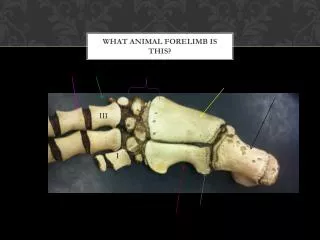
Dog Elbow Q3 • (i) What is 1? • Anconeal Process • Olecranon Process • Medial Coronoid Process • (ii) What is 2? • Olecranon Process • Medial Coronoid Process • Anconeal Process • (iii) Structure 2 articulates with which part of 7? • Olecranon Fossa • Supracondyloid Foramen • (iv) What is 5? • Radius • Ulna • (v) What is 6? • Radius • Ulna 6 7 5
Correct • Well done! (1) is the Olecranon process! • Remember: the Olecranon Process is the point of insertion for the Triceps muscle, the main extensor muscle of the elbow. • Here are more examples • Try (ii)! • Choose a new question. Olecranon Olecranon
Incorrect • No, (1) is not the Anconeal Process. • Here are x-rays showing the Anconeal Process . • Try again! • Choose a new question. Anconeal Process Anconeal Process
Incorrect • No, (1) is not the Medial Coronoid Process. • These x-rays show the Medial Coronoid Process. • Try again! • Choose a new question. Coronoid Process Coronoid Process
Correct • Well done! (2) is the Anconeal Process! • Remember: the Anconeal Process articulates with the Olecranon Fossa of the Humerus • Here are some more examples. • Try (iii)! • Choose a new question. Anconeal Process Anconeal Process
Incorrect • No, (2) is not the Olecranon process. • Here are some x-rays showing the Olecranon process. • Try again! • Choose a new question. Olecranon Olecranon
Incorrect • No, (2) is not the Medial Coronoid Process. • Remember: the Medial and Lateral Coronoid Processes of the radius are the points of attachment for the annular ligament that holds the ulna in place. • The Coronoid Process is labelled in these x-rays. • Try again! • Choose a new question. Coronoid Process Coronoid Process
Correct • Well done! • The Anconeal Process of the Ulna articulates with the Olecranon Fossa of the Humerus! • Try (iv)! • Choose a new question. Olecranon Fossa Anconeal Process
Incorrect • No, the Anconeal Processdoes not articulate with the Supracondyloid Foramen. • This x-ray shows the Supracondyloid foramen • Remember: this is present in the cat but not the dog and blood vessels pass through it. • Try again! • Choose a new question. Supracondyloid Foramen
Correct • Yes! (5) is the Ulna! • Remember: the Ulna has the large Olecranon Process proximally and tapers distally in the dog. • Here are more x-rays of the Ulna. • Try (v)! • Choose a new question. Ulna Ulna
Incorrect Radius • No, (5) is not the Radius. • Here are some x-rays of the Radius. • Try again! • Choose a new question. Radius
Correct Radius • Yes! (6) is the Radius! • Remember: the Radius is the main weight bearing bone in the antebrachium • Here are more x-rays of the radius. • Try Dog Elbow Q2! • Choose a new question. Radius
Incorrect • No, (6) is not the Ulna. • Here are some x-rays of the Ulna. • Try again! • Choose a new question. Ulna Ulna
Dog Elbow Q4 • (i) What is 7? • Medial Coronoid Process • Medial Epicondyle • Lateral Epicondyle • (ii) What is 8? • Anconeal process • Olecranon process • Head of Radius • (iii) What is 12? • Distal humeral growth plate • Supracondyloid Foramen • Olecranon Fossa
Correct • Yes! (7) is the Medial Coronoid Process! • Remember: the Medial and Lateral Coronoid Processes of the radius are the points of attachment for the annular ligament that holds the ulna in place. • Here are other x-rays showing the Medial Coronoid Process. • Try (ii)! • Choose a new question. Coronoid Process What can you say about the age of this animal? Answer. Coronoid Process
Answer • This is a young dog! • Notice the growth plates – they are particularly obvious at the proximal end of the radius. • Go Back!
Incorrect • No, (7) is not the Medial Epicondyle. • This x-ray shows the Medial Epicondyle. • Try again! • Choose a new question. Medial Epicondyle
Incorrect • No, (7) is not the Lateral Epicondyle. • This x-ray shows the Lateral Epicondyle. • Try again! • Choose a new question. Lateral Epicondyle
Correct • Yes! (8) is the Olecranon process! • Here are more examples. • Try (iii)! • Choose a new question. Olecranon Olecranon
Incorrect • No, (8) is not the Anconeal process. • These x-rays show the Anconeal process. • Try again! • Choose a new question. Anconeal process Anconeal process
Incorrect Head of Radius • No, (8) is not the Head of the Radius. • These x-rays show the Head of the Radius. • Try again! • Choose a new question. Head of Radius
Correct • Yes! (12) is the Olecranon Fossa! • Here is another example. • Try Dog Carpus Q1. • Choose a new question. Olecranon Fossa
Incorrect • No, (12) is not the Distal Humeral growth plate / physis. • This x-ray shows the Distal Humeral growth plate / physis. • Try again! • Choose a new question. Distal humeral growth plate