Basic radiation protection & radiobiology
230 likes | 780 Vues
Basic radiation protection & radiobiology. By Dr. Mohsen Dashti Patient care & management 202 14-3-10. Discussion issues. Ionizing radiation. Protecting the patient. Protecting the radiographer. Radiation monitoring. . Ionizing radiation. What are the sources of ionizing radiation?

Basic radiation protection & radiobiology
E N D
Presentation Transcript
Basic radiation protection & radiobiology By Dr. Mohsen Dashti Patient care & management 202 14-3-10
Discussion issues • Ionizing radiation. • Protecting the patient. • Protecting the radiographer. • Radiation monitoring.
Ionizing radiation • What are the sources of ionizing radiation? • Natural radiation. - What is natural radiation? -- Sources of radiation that occur spontaneously in nature and can be affected by human activity. • Examples: -- Cosmic radiation….. The sun and other planets. -- Radioactive substances on earth…. Uranium and radium. • Natural radiation sources are given less attention to their hazardous potential.
Ionizing radiation • What are the sources of ionizing radiation? • Manmade radiation. - What is manmade radiation? -- Sources of radiation that are developed by humans and used in different fields of technology. • Examples: -- Nuclear industry…. Weapons & nuclear power stations. -- Radionuclide…. Radioactive elements & radiopharmaceuticals. -- Medical radiation…. Medical imaging & dental exposure.
Ionizing radiation • Manmade radiation. • It is known as x-rays, which is a form of electromagnetic radiation that travels at the speed of light depositing energy randomly. • How can we produce x-rays? • Source of electrons. • Force to move electrons rapidly. • Element to stop this movement rapidly.
Ionizing radiation • Manmade radiation. • What happens to x-rays when they are produced? • Absorbed. • Scatter. • Pass through undistributed.
Ionizing radiation • Manmade radiation. • How do x-rays interact with matter? • Classic coherent scattering. -- Interaction with matter in which a low-energy photon (below 10 keV) is absorbed and released with its same energy, frequency and wavelength but with change of direction. • Photoelectric interaction. -- Interaction with matter in which proton strikes an inner shell electron, causing its ejection from orbit with complete absorption of the photon’s energy.
Ionizing radiation • Manmade radiation. • How do x-rays interact with matter? • Compton scattering. -- Interaction with matter in which a higher-energy photon strikes a loosely bound outer electron, removing it from its shell, and the remaining energy is released as scatter photon. • Pair production. -- Interaction between matter and photon possessing a minimum of 1.02 MeV of energy, producing two oppositely charged particles. • Photodisintegration. -- Interaction directly with the nucleus of photon possessing a minimum of 10 MeV, causing excitement followed by emission of nuclear fragment.
Ionizing Radiation • Standards for regulation of exposure: • What guidelines available to limit radiation dose? • No-threshold. -- No dose exists below which the risk of damage does not exist. 2. Risk versus benefit. -- The benefit to the patient performing radiographic procedure far outweigh the risk of possible biologic damage.
Ionizing radiation • Radiation risk.
Ionizing radiation • ALARA… • To keep radiation dose as low as reasonably achievable. -- The annual whole-body dose-equivalent limit for the occupational worker is 50mSv (5 rem). -- The whole-body dose-equivalent limit for the general population is one tenth the occupational worker’s annual limit or 5 msv (0.5 rem). • Sv: unit in the SI system to measure the dose-equivalent or biologic effectiveness of differing radiation; 1 Sv is equal to 100 rems.
Protecting the patient • ALARA concept can be practiced with the patient by utilizing 3 methods: • Time: • Time minimization is the most important element to protect the patient from radiation dose. How? -- Applying the rules of radiographic techniques. -- Using the exposure chart to determine the correct amount of radiation to produce an image. -- Minimizing repeat rates to reduce the patient’s time in the path of the x-ray beam.
Protecting the patient • Distance: • Distance maximization is another element to reduce patient radiation dose. Why? -- This serve to lessens the skin or entrance dose to the patient. -- Increasing the distance should be kept to a reasonable range so radiation dose will not be affected. How? -- For you to answer??? • Shielding: • Use of shield to protect sensitive or unexposed region of the patient’s body is another method to protect the patient from radiation dose.
Protecting the patient • Shielding: • The rule indicates that patients should be shielded whenever they are 4-5 cm from the primary x-ray beam. -- Shields are made of lead, which absorbs x-rays through the process of photoelectric effect, thereby minimizing patient exposure. • Types of shield: • Flat contact shield: made of a combination of vinyl and lead. Placed directly over the gonads of the patient. • Shaped shield: cup shaped and made specifically for male patients.
Protecting the patient • Shadow shield: mounted on the side of the collimator of the x-ray tube and can be manipulated to extend into the path of the beam.
Protecting the radiographer • The same methods are used to protect the radiographer from extra radiation dose. • The radiographer should spend the least amount of time possible in a room when a source of radiation is active. • Fluoroscopy requires the radiographer to spend longer time in an active radiation room, therefore extra protection should be considered. • Distance is the best measure to protect the radiographer from radiation dose. • Inverse square law should be applied to reduce the impact of radiation dose.
Protecting the radiographer • Inverse square law: The intensity of radiation varies inversely with the square of the distance. What does it mean? -- For you to answer??? • Submit your answer next week
Protecting the patient • Lead shield and aprons must be used by the radiographer whenever radiation is active. • Aprons and lead shields must in in good conditions and crack free to avoid passing radiation into the radiographer. • The minimum permissible amount of lead equivalency for aprons used where the peak kilovoltage is 100 should be 0.25 mm.
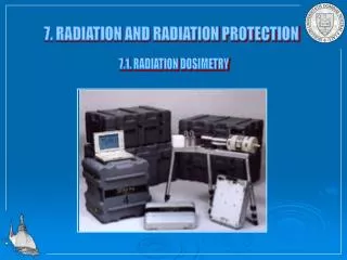
Radiation monitoring • Discuss the four main radiation monitoring methods used in x-rays; film badges, thermoluminescent dosimeters, pocket dosimeters, and field survey instruments.