
The Respiratory System
The Respiratory System. Respiration Includes. Pulmonary ventilation Air moves in and out of lungs Continuous replacement of gases in alveoli (air sacs) External respiration Gas exchange between blood and air at alveoli O2 (oxygen) in air diffuses into blood

The Respiratory System
E N D
Presentation Transcript
Respiration Includes • Pulmonary ventilation • Air moves in and out of lungs • Continuous replacement of gases in alveoli (air sacs) • External respiration • Gas exchange between blood and air at alveoli • O2 (oxygen) in air diffuses into blood • CO2 (carbon dioxide) in blood diffuses into air • Transport of respiratory gases • Between the lungs and the cells of the body • Performed by the cardiovascular system • Blood is the transporting fluid • Internal respiration • Gas exchange in capillaries between blood and tissue cells • O2 in blood diffuses into tissues • CO2 waste in tissues diffuses into blood
Cellular Respiration • Oxygen (O2) is used by the cells • O2 needed in conversion of glucose to cellular energy (ATP) • All body cells • Carbon dioxide (CO2) is produced as a waste product • The body’s cells die if either the respiratory or cardiovascular system fails
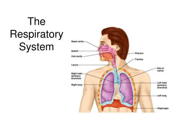
The Respiratory Organs Conducting zone • Respiratory passages that carry air to the site of gas exchange • Filters, humidifies and warms air Respiratory zone • Site of gas exchange • Composed of • Respiratory bronchioles • Alveolar ducts • Alveolar sacs Conducting zone labeled
Conducting zone will be covered first Nose • Provides airway • Moistens and warms air • Filters air • Resonating chamber for speech • Olfactory receptors External nose
Linings of nasal cavity • Vestibule* (just above nostrils) • Lined with skin containing sebaceous and sweat glands and nose hairs • Filters large particulars (insects, lint, etc.) • The remainder of nasal cavity: 2 types of mucous membrane • Small patch of olfactory mucosa near roof (cribriform plate) • Respiratory mucosa: lines most of the cavity Olfactory mucosa *
Paranasal sinuses • Frontal, sphenoid, ethmoid and maxillary bones • Open into nasal cavity • Lined by same mucosa as nasal cavity and perform same functions • Also lighten the skull • Can get infected: sinusitis
The Pharynx (throat) • 3 parts: naso-, oro- and laryngopharynx • Houses tonsils (they respond to inhaled antigens) • Uvula closes off nasopharynx during swallowing so food doesn’t go into nose • Epiglottis posterior to the tongue: keeps food out of airway • Oropharynx and laryngopharynx serve as common passageway for food and air • Lined with stratified squamous epithelium for protection * *
The Larynx (voicebox) • Three functions: • Produces vocalizations (speech) • Provides an open airway (breathing) • Switching mechanism to route air and food into proper channels • Closed during swallowing • Open during breathing
* * Posterior views Epliglottis* (the 9th cartilage) Elastic cartilage covered by mucosa On a stalk attached to thyroid cartilage Attaches to back of tongue During swallowing, larynx is pulled superiorly Keeps food out of lower respiratory tract
Glottis is the space between the vocal cords • Laryngeal muscles control length and size of opening by moving arytenoid cartilages • Sound is produced by the vibration of vocal cords as air is exhaled
Trachea (the windpipe) • Descends: larynx through neck into mediastinum • Divides in thorax into two main (primary) bronchi • Flexible for bending but stays open despite pressure changes during breathing
Respiratory Zone • Structures that contain air-exchange chambers are called alveoli • Respiratory bronchioles lead into alveolar ducts: walls consist of alveoli • Ducts lead into terminal clusters called alveolar sacs – are microscopic chambers • There are 3 million alveoli!
Gas Exchange • Air filled alveoli account for most of the lung volume • Very great area for gas exchange (1500 sq ft) • Alveolar wall • Single layer of squamous epithelial cells Respiratory bronchiole Alveolar duct (air on one side; blood on the other) Alveoli Alveolar sac
Bronchial “tree” and associated Pulmonary arteries
This “air-blood barrier” (the respiratory membrane) is where gas exchange occurs • Oxygen diffuses from air in alveolus (singular of alveoli) to blood in capillary • Carbon dioxide diffuses from the blood in the capillary into the air in the alveolus
Surfactant • Type II cuboidal epithelial cells are scattered in alveolar walls • Surfactant is a detergent-like substance which is secreted in fluid coating alveolar surfaces – it decreases tension • Without it the walls would stick together during exhalation • Premature babies – problem breathing is largely because lack surfactant
Lungs and Pleura Around each lung is a flattened sac of serous membrane called pleura Parietal pleura – outer layer Visceral pleura – directly on lung Pleural cavity – slit-like potential space filled with pleural fluid • Lungs can slide but separation from pleura is resisted (like film between 2 plates of glass) • Lungs cling to thoracic wall and are forced to expand and recoil as volume of thoracic cavity changes during breathing
CXR (chest x-ray)
Lungs • Each is cone-shaped with anterior, lateral and posterior surfaces contacting ribs • Superior tip is apex, just deep to clavicle • Concave inferior surface resting on diaphragm is the base apex apex base base
Abbreviations in medicine: e.g.” RLL pneumonia” Horizontal fissure • Right lung: 3 lobes • Upper lobe • Middle lobe • Lower lobe • Left lung: 2 lobes • Upper lobe • Lower lobe Oblique fissure Oblique fissure Each lobe is served by a lobar (secondary) bronchus
Ventilation • Breathing = “pulmonary ventilation” • Pulmonary means related to the lungs • Two phases • Inspiration (inhalation) – air in • Expiration (exhalation) – air out • Mechanical forces cause the movement of air • Gases always flow from higher pressure to lower • For air to enter the thorax, the pressure of the air in it has to be lower than atmospheric pressure • Making the volume of the thorax larger means the air inside it is under less pressure (the air has more space for as many gas particles, therefore it is under less pressure) • The diaphragm and intercostal muscles accomplish this
Muscles of Inspiration • During inspiration, the dome shaped diaphragm flattens as it contracts • This increases the height of the thoracic cavity • The external intercostalmuscles contract to raise the ribs • This increases the circumference of the thoracic cavity Together:
Expiration • Quiet expiration in healthy people is chiefly passive • Inspiratory muscles relax • Rib cage drops under force of gravity • Relaxing diaphragm moves superiorly (up) • Elastic fibers in lung recoil • Volumes of thorax and lungs decrease simultaneously, increasing the pressure • Air is forced out
Expiration continued • Forced expiration is active • Contraction of abdominal wall muscles • Oblique and transversus predominantly • Increases intra-abdominal pressure forcing the diaphragm superiorly • Depressing the rib cage, decreases thoracic volume • Some help from internal intercostals and latissimus dorsi (try this on yourself to feel the different muscles acting)
Pneumothorax (collapsed lung) • Think about the processes involved and then try and imagine the various scenarios • Trauma causing the thoracic wall to be pierced so air gets into the pleura • Broken rib can do (1); always do a CXR if there’s a broken rib • Visceral pleura breaks, letting alveolar air into pleural space
There are many diseases of the respiratory system, including asthma, cystic fibrosis, COPD (chronic obstructive pulmonary disease – with chronic bronchitis and/or emphysema) and epiglottitis example: normal emphysema


