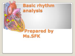
Basic rhythm analysis Prepared by Ms.SFK
Basic rhythm analysis Prepared by Ms.SFK. By the end of the presentation learners will be able to Define the term electrocardiography, electrocardiogram, and electrocardiograph Explore placement of ECG electrodes Develop understanding about ECG graph paper

Basic rhythm analysis Prepared by Ms.SFK
E N D
Presentation Transcript
By the end of the presentation learners will be able to • Define the term electrocardiography, electrocardiogram, and electrocardiograph • Explore placement of ECG electrodes • Develop understanding about ECG graph paper • Identify relationship between conducting system and PQRST complex • Discuss steps of reading rhythm strips Objectives
Conduction System and PQRST Complex Ventricular depolarization Ventricular contraction Ventricular diastole Atrial depolarization Atrial contraction Atrial diastole
Conduction System and PQRST Complex Atrial rate 60-80 Below ventricles 20-40 Ventricular rate 40-60
Represents atrial depolarization. • Usually 2.5mm or less in height and 0.11second or less in duration. • Upright P Wave
Represents ventricular depolarization • Q wave is first negative deflection and less than 0.03 sec and less than 25% of R wave amplitude. • R wave is first positive deflection • S wave is first negative deflection after R wave • QRS duration is 0.04-0.10secs QRS
Ventricular re polarization. • U wave may indicate electrolyte abnormality. It is thought to be result of slow re polarization of intra ventricular conduction system. T and U waves
Represents early re polarization of the ventricles PR interval • Represents time required for the impulse to travel from atria and conduction system to purkkinje fibers. • PR interval ranges from 0.12-0.20 secs. ST segment
Represents electrical systole • Is usually half of the RR interval • 0.32-0.40 sec if heart rate is 65-95min. • QT interval varies with heart rate therefore QTc should be calculated. • QTc should not exceed 0.42 sec in men and 0.43 sec in women. QT interval
Systematic approach to rhythm strip analysis • Rate (atrial and ventricular rate) • Rhythm (PP.RR interval) • Interval • PR • QRS • QT
Determination of Heart rate • 6 sec method • Count down method
PP or RR interval Determination of rhythm
SA Node • Atria • AV node/ junction • Ventricles Arrhythmias
Normal Sinus Rhythm • Rate (60-100/min) atrial and ventricular rates are equal and every p has QRS. • Rhythm (regular) • Intervals normal Dysrhythmias of SA nodes
Sinus Tachycardia HR 100-160
PAC (Premature atrial contraction Earlier than expected P waves — morphology of premature P wave usually differs from the sinus P. • Atrial P wave often falls near the T wave and distorts it — P is flat, notched, or lost in the T in at least one lead. • PR interval is usually normal. • Rhythm is regular except for the premature contraction
Impulse from atrial ectopic foci P wave sawtoothed, QRS complex narrow, atrial rate 250-350, Atrial Flutter
Atrial and ventricular rate and rhythm are irregular Atrial rate 350-500 Multiple impulses generate from atria (ectopic foci) Atrial Fibrillation
In this arrhythmia, atrial tachycardia occurs with numerous atrial foci firing intermittently. • MAT produces varying P waves on the Strip and occurs most often in patients with chronic pulmonary disease Multifocal Atrial Tachycardia (MAT)
Atrial Rate: 100 to 250 beats/minute • Ventricular Rate: > 160 • P wave: Three different Shapes • Rhythm: Both irregular • PR interval: Varies