Spinal Cord and Spinal Nerves
901 likes | 2.41k Vues
Chapter 12. Spinal Cord and Spinal Nerves. Spinal Meninges. Connective tissue membranes surrounding spinal cord and brain Dura mater : continuous with epineurium of the spinal nerves----thick Arachnoid mater : thin and wispy Pia mater : bound tightly to surface of brain and spinal cord. .

Spinal Cord and Spinal Nerves
E N D
Presentation Transcript
Chapter 12 Spinal Cord and Spinal Nerves
Spinal Meninges • Connective tissue membranes surrounding spinal cord and brain • Dura mater: continuous with epineurium of the spinal nerves----thick • Arachnoid mater: thin and wispy • Pia mater: bound tightly to surface of brain and spinal cord.
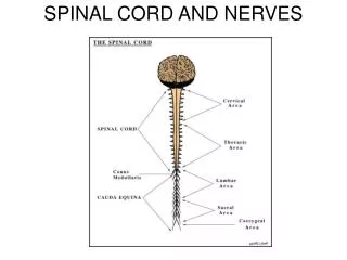
Cross Section of Spinal Cord • Roots: spinal nerves arise as rootlets then combine to form roots • Dorsal (posterior) root has a ganglion • Ventral (anterior) • Two roots merge laterally and form the spinal nerve
Organization of Neurons in the Spinal Cord and Spinal Nerves • Dorsal root ganglion: collections of cell bodies of unipolar sensory neurons forming dorsal roots. • Motor neuron cell bodies are in anterior and lateral horns of spinal cord gray matter. • Multipolar somatic motor neurons in anterior (motor) horn • Autonomic neurons in lateral horn • Axons of motor neurons form ventral roots and pass into spinal nerves
Reflex Arc • Basic functional unit of nervous system and simplest portion capable of receiving a stimulus and producing a response • Automatic response to a stimulus that occurs without conscious thought. Homeostatic. • Components • Action potentials produced in sensory receptors transmitted to • Sensory neuron. To-Interneurons. To-Motor neuron. To- • Effector organ which responds with a reflex
Structure of Peripheral Nerves • Consist of • Axon bundles • Schwann cells • Connective tissue • Endoneurium: surrounds individual neurons • Perineurium: surrounds axon groups to form fascicles • Epineurium: surrounds the entire nerve
Branches of Spinal Nerves • Dorsal Ramus: innervate deep muscles of the trunk responsible for movements of the vertebral column and the C.T. and skin near the midline of the back. • Ventral Ramus: what they innervate depends upon which part of the spinal cord is considered. • Thoracic region: form intercostal nerves that innervate the intercostal muscles and the skin over the thorax • Remaining spinal nerve ventral rami (roots of the plexus): form five plexuses (intermingling of nerves). • Ventral rami of C1-C4= cervical plexus • Ventral rami of C5-T1= brachial plexus • Ventral rami of L1-L4= lumbar plexus • Ventral rami of L4-S4= sacral plexus • Ventral rami of S4 and S5= coccygeal plexus
Chapter 13 Brain and Cranial Nerves
Brain and Cranial Nerves • Brain • Part of CNS contained in cranial cavity • Control center for many of body’s functions • Much like a complex computer but more • Parts of the brain • Brainstem: connects spinal cord to brain; integration of reflexes necessary for survival • Cerebellum: involved in control of locomotion, balance, posture • Diencephalon: thalamus, hypothalamus • Cerebrum: conscious thought, control • Cranial nerves: part of PNS arise directly from brain. Two pairs arise from cerebrum; ten pairs arise from brainstem
Brainstem: Medulla Oblongata • Most inferior part • Continuous with spinal cord; has both ascending and descending nerve tracts • Regulates: heart rate, blood vessel diameter, respiration, swallowing, vomiting, hiccupping, coughing, and sneezing Brainstem: Pons • Superior to the medulla oblongata • Sleep center • Respiratory center coordinates with center in medulla
Brainstem: Midbrain • Also called mesencephalon • Small and superior to pons • Tectum: four nuclei that form mounds on dorsal surface of midbrain. Corpora quadrigemina • Each separate part is a colliculus • Two superior colliculi involved in visual reflexes; receive information from inferior colliculi, eyes, skin, cerebrum • Two inferior colliculi involved in hearing
ReticularFormation • Group of nuclei scattered throughout brainstem • Controls cyclic activities such as sleep-wake cycle (maintains alertness)
Cerebellum • Attached to brainstem posterior to pons • Cerebellar peduncles: fiber tracts that communicate with other parts of brain • Superior: to midbrain • Middle: to pons • Inferior: to medulla oblongata • Gray cortex and nuclei with white matter (tracts) between • Cortex folded in ridges called folia; white matter resembles a tree (arbor vitae)
Cerebellar Functions • Flocculonodular lobe: balance and eye movements • Vermis and medial portion of hemispheres: posture, locomotion, fine motor coordination leading to smooth, flowing movements • Lateral hemispheres, major portion: works with cerebrum to plan, practice, learn complex movements
Diencephalon • Located between brainstem and cerebrum • Components: thalamus, subthalamus, epithalamus, hypothalamus
Thalamus • Two lateral portions connected by the intermediate mass • Surrounded by third ventricle • Sensory information from spinal cord synapses here before projecting to cerebrum • Medial geniculate nucleus: auditory information • Lateral geniculate nucleus: visual information • Ventral posterior nucleus: most other types sensory information
Hypothalamus • Most inferior portion of diencephalon • Mammilary bodies: bulges on ventralsurface; olfactory reflexes and emotional responses to odors • Infundibulum: stalk extending from floor; connects hypothalamus to posterior pituitary gland. Controls endocrine system. • Receives input from viscera, taste receptors, nipples, external genitalia, prefrontal cortex • Efferent fibers to brainstem, spinal cord (autonomic system), through infundibulum to posterior pituitary, and to cranial nerves controlling swallowing and shivering • Important in regulation of mood, emotion,sexual pleasure, satiation, rage, and fear
Cerebrum • Largest portion of brain • Composed of right and left hemispheres each of which has the following lobes: frontal, parietal, occipital, temporal • Sulci and Fissures • Longitudinal fissure: separates the two hemispheres • Lateral fissure: separates temporal lobe from frontal and parietal lobes • Central sulcus: separates frontal and parietal lobes • Cortex: outer surface • Gyri are folds (increase surface area) • Sulci are depressions
Cerebrum, cont. • Central sulcus: between the precentral gyrus (primary motor cortex) and postcentral gyrus (primary somatic sensory cortex) • Frontal lobe: voluntary motor function, motivation, aggression, sense of smell, mood • Parietal lobe: reception and evaluation of sensory information except smell, hearing, and vision • Occipital lobe: reception and integration of visual input • Temporal lobe: reception and evaluation for smell and hearing; memory, abstract thought, judgment. Insula is within.
Meninges • Connective tissue membranes • Dura mater: superficial • Arachnoid mater • Pia mater: bound tightly to brain • Spaces • Subdural: serous fluid • Subarachnoid: CSF
Ventricles • Lateral ventricles: within cerebral hemispheres; separated by • septa pellucida • Third ventricle: within diencephalon • Interventricular foramina join lateral ventricles with third • Fourth ventricle: associated with pons and medulla oblongata. Connected to third ventricle by the cerebral aqueduct, continuous with the spinal cord.
Cerebrospinal Fluid (CSF) • Similar to serum, but most protein removed • Bathes brain and spinal cord • Protective cushion around CNS • Choroid plexuses produce CSF which fills ventricles and other parts of brain and spinal cord • Composed of ependymal cells, their support tissue, and associated blood vessels • Blood-cerebrospinal fluid barrier • Endothelial cells of capillaries attached by tight junctions • Substances do not pass between cells • Substances must pass through cells • Makes the barrier very selective
Integration of Nervous System Functions Chapter 14
Senses • Means by which brain receives information about environment and body. • Sensation (perception): conscious awareness of stimuli received by sensory receptors. • Steps to sensation • Stimuli originating either inside or outside of the body must be detected by sensory receptors and converted into action potentials, which are propagated to the CNS by nerves. • Within the CNS, nerve tracts convey action potentials to the cerebral cortex and to other areas of the CNS. • Action potentials reaching the cerebral cortex must be translated so the person can be aware of the stimulus.
Types of Senses • General: distributed over large part of body. Receptor generates an action potential called a generator potential that then travels to the brain. Called primary receptors. • Somatic (information about the body and environment): touch, pressure, temperature, proprioception, pain • Visceral (information about internal organs): pain and pressure • Special senses: smell, taste, sight, hearing, balance.
Types of Sensory Receptors • Mechanoreceptors: compression, bending, stretching of cells. Touch, pressure, proprioception, hearing, and balance • Chemoreceptors: chemicals become attached to receptors on their membranes. Smell and taste • Thermoreceptors: respond to changes in temperature • Photoreceptors: respond to light: vision • Nociceptors: extreme mechanical, chemical, or thermal stimuli. Pain
Types of Receptors Based on Location • Exteroreceptors: associated with skin • Visceroreceptors: associated with organs • Proprioceptors: associated with joints, tendons (proprioception is the sense of the orientation of one's limbs in space)
Free Nerve Endings • Simplest, most common sensory receptor • Scattered through most of body; visceroceptors are of this type. • Type responsible for temperature sensation • Cold: 10-15 times more numerous than warm • Warm • Pain: responds to extreme cold or heat
Merkel (Tactile) Disks • Associated with dome-shaped mounds of thickened epidermis in hairy skin • Light touch and superficial pressure
Pacinian Corpuscles • Deep cutaneous pressure; vibration • When associated with joints, involved in proprioception (proprioception is the sense of the orientation of one's limbs in space)
Meissner (Tactile) Corpuscles • Two-point discrimination • Ability to detect simultaneous stimulations at two points on the skin. • Used to determined texture of objects. • Numerous and close together on tongue and fingertips • Light touch & light pressure
Sensory and Association Areasof the Cerebral Cortex • Sensory • Primary somatic sensory cortex (general sensory area): posterior to the central sulcus. Postcentral gyrus. • General sensory input: pain, pressure, temperature • Taste area: inferior end of postcentral gyrus • Olfactory cortex: inferior surface of frontal lobe • Primary auditory cortex: superior part of temporal lobe • Visual cortex: occipital lobe • Association areas: process of recognition • Somatic sensory: posterior to • primary somatic sensory cortex • Visual association: anterior to • visual cortex: present visual • information compared to past • information
Referred Pain • Referred: sensation in one region of body that is not source of stimulus. Organ pain usually referred to the skin. Both the organ and that region of the skin input to the same spinal segment and converge on the same ascending neurons.
Phantom and Chronic Pain • Phantom: occurs in people who have appendage amputated or structure removed such as a tooth. Gate control theory of pain-- in uninjured limb, pressure and touch sensation inhibits pain (thus the success of massage in pain relief). These sensations are lost with amputations and thus their inhibitory effect.
Control of Skeletal Muscles • Motor system: maintains posture and balance; moves limbs, trunk, head, eyes; facial expression, speech. • Reflexes: movements that occur without conscious thought • Voluntary movements: consciously activated to achieve a specific goal • Two neurons: upper and lower • Upper motor neurons: directly or through interneurons connect to lower • Lower motor neurons: axons leave the CNS, extend through PNS to skeletal muscles. Cell bodies in anterior horns of spinal cord and in cranial nerve nuclei of brainstem
Motor Areas of the Cerebral Cortex • Precentral gyrus (primary motor cortex, primary motor area): 30% of upper motor neurons. Another 30% in premotor area. Control voluntary movements, especially fine motor movements of hands • Premotor area: anterior to primary motor cortex. Motor functions organized before initiation • Prefrontal area: motivation, foresight to plan and initiate movements, emotional behavior, mood
Direct Pathways • Control muscle tone and conscious fine, skilled movements in the face and distal limbs • Direct synapse of upper motor neurons of cerebral cortex with lower motor neurons in brainstem or spinal cord • Tracts • Corticospinal: direct control of movements below the head • Corticobulbar: direct control of movements in head and neck
Indirect Pathways • Control conscious and unconscious muscle movements in trunk and proximal limbs. • Synapse in some intermediate nucleus rather than directly with lower motor neurons. • Tracts • Rubrospinal: upper neurons synapse in red nucleus. Similar to comparator function of cerebellum. Regulates fine motor control of muscles in distal part of upper limb. • Vestibulospinal: influence neurons innervating extensor muscles in trunk and proximal portion of lower limbs; help maintain upright posture. • Reticulospinal: maintenance of posture.
Cerebellum • Helps maintain muscle tone in postural muscles, helps control balance during movement, and coordinate eye movements
Cerebellar Comparator Function • The motor cortex sends action potentials to lower motor neurons in the spinal cord. • Action potentials from the motor cortex inform the cerebellum of the intended movement. • Lower motor neurons in the spinal cord send action potentials to skeletal muscles, causing them to contract. • Proprioceptive signals from the skeletal muscles and joints to the cerebellum convey information & cerebellum helps accomplish fine motor coordination of simple movements. It compares the intended movement with the actual movement and the result is smooth & coordinated movements. (ex: touching your nose--------not rapid complex movements)
Speech • Area normally in left cerebral cortex • Wernicke's area: sensory speech- understanding what is heard and thinking of what one will say. • Broca's area: motor speech- sending messages to the appropriate muscles to actually make the sounds. • Sound is heard first in the 1o association area, then information travels to Wernicke's area. Neuronal connections between Wernicke's area and Broca's area. • Aphasia: absent or defective speech or language comprehension. Caused by lesion somewhere in the auditory/speech pathway.
Right and Left Cerebral Cortex • Right: controls muscular activity in and receives sensory information from left side of body • Left: controls muscular activity in and receives sensory information from right side of body • Sensory information of both hemispheres shared through commissures: corpus callosum • Language, and possibly other functions like artistic activities, not shared equally • Left: mathematics and speech • Right: three-dimensional or spatial perception, recognition of faces, musical ability
Chapter 16 Autonomic Nervous System
Peripheral Nervous System • Peripheral nerves contain both motor and sensory neurons • Among the motor neurons, some of these are somatic and innervate skeletal muscles while some are autonomic and innervate smooth muscle, cardiac muscle, and glands • Sensory neurons are not subdivided into somatic and autonomic since there is overlap in function; e.g., pain receptors can stimulate both somatic and autonomic reflexes
Somatic Skeletal muscle Conscious and unconscious movement Skeletal muscle contracts One synapse Acetylcholine Receptor molecules: nicotinic Autonomic Smooth and cardiac muscle and glands Unconscious regulation Target tissues stimulated or inhibited Two synapses Acetylcholine by preganglionic neurons and ACh or norepinephrine by postganglionic neurons Receptor molecules: varies with synapse and neurotransmitter Somatic and Autonomic Nervous Systems