Spinal Cord, Spinal Nerves, and Somatic Reflexes
170 likes | 627 Vues
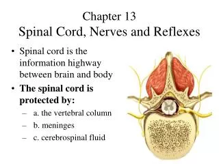
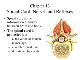
Functions of the Spinal Cord. Conduction ? sends information up and down the cordLocomotionReflexes ? involuntary responses to stimuli. Anatomy of the Spinal Cord. Occupies C1 through L1 ? starting at the foramen magnumCauda equina Filum terminaleDivided into 4 regionsCervicalThoracicLumbar

Spinal Cord, Spinal Nerves, and Somatic Reflexes
E N D
Presentation Transcript
1. Spinal Cord, Spinal Nerves, and Somatic Reflexes
2. Functions of the Spinal Cord Conduction � sends information up and down the cord
Locomotion
Reflexes � involuntary responses to stimuli
3. Anatomy of the Spinal Cord Occupies C1 through L1 � starting at the foramen magnum
Cauda equina
Filum terminale
Divided into 4 regions
Cervical
Thoracic
Lumbar
Sacral Cauda equina - Occupies the vertebral canal from L2 to S5 � bundle of nerve roots
Nerves named for the levels of the vertebral column through which the spinal nerves emerge
Cervical and lumbar are widest regions � for the emergence of nerves that control the limbsCauda equina - Occupies the vertebral canal from L2 to S5 � bundle of nerve roots
Nerves named for the levels of the vertebral column through which the spinal nerves emerge
Cervical and lumbar are widest regions � for the emergence of nerves that control the limbs
4. Meninges Enclose the spinal cord
Three layers
Dura mater � superficial
Arachnoid mater
Pia mater � deep
Space between
Epidural � between dura mater and vertebral bone
Subarachnoid � between arachnoid and pia mater Dura mater � tough collagenous membrane
Epidural space between � has blood vessels, adipose tissue, and connective tissue � anesthetics are introduced to this space to block pain signals � an epidural
Arachnoid mater � loose mesh of collagenous and elastic fibers
Subarachnoid � filled with CSF
Pia mater � delicate, translucent membraneDura mater � tough collagenous membrane
Epidural space between � has blood vessels, adipose tissue, and connective tissue � anesthetics are introduced to this space to block pain signals � an epidural
Arachnoid mater � loose mesh of collagenous and elastic fibers
Subarachnoid � filled with CSF
Pia mater � delicate, translucent membrane
5. Nervous tissue Gray matter
Contains somas, dendrites, and synapses
Little myelin
Site of information processing
White matter
Contains nerve fibers (axons)
Abundance of myelin
Carry signals from one part
of the CNS to another
Information processing in CNSInformation processing in CNS
6. Gray Matter Looks like a butterfly
2 dorsal horns
2 ventral horns
Central canal
Spinal nerve branches into a dorsal root and ventral root
Dorsal root
Ventral root Dorsal horns are posterior
Ventral horns are anterior
DORSAL root � carries sensory nerve fibers
VENTRAL horns � contain large somas of the somatic morotr neurons � axons exit by way of the ventral root of the spinal nerve and lead to the skeletal muscleDorsal horns are posterior
Ventral horns are anterior
DORSAL root � carries sensory nerve fibers
VENTRAL horns � contain large somas of the somatic morotr neurons � axons exit by way of the ventral root of the spinal nerve and lead to the skeletal muscle
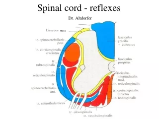
8. White Matter Surrounds the gray matter
Bundles of axons that provide avenues of communication up and down the spinal cord
Bundles are arranged in three pairs of columns
Dorsal
Lateral
Ventral
Each column has subdivisions called tracts
9. Spinal Tracts Ascending �
Travel across three neurons from their origin in the receptors to their destination in the sensory areas of the
First-order neuron � detects stimuli and sends it to the spinal cord or brainstem
Second-order � continues to the thalamus at the upper end of the brainstem
Third-order � carries the signal to the sensory region of the cerebral cortex
carry sensory information up the cord
Braincarry sensory information up the cord
Brain
10. Spinal Tracts Descending �
Two neurons
Upper motor neuron � begins with a soma in the cerebral cortex or brainstem
Lower motor neuron � where the axon terminates from the above mentioned soma and travels to the muscle or target organ DESCENDING � carry motor impulses down the brainstem and spinal cord
DESCENDING � carry motor impulses down the brainstem and spinal cord
11. Spinal Nerves 31 pairs
8 cervical
12 thoracic
5 lumbar
1 coccygeal
Spinal nerves are mixed nerves � carries signals both ways
Afferent signals approach the cord by the dorsal root and enter the dorsal horn of the gray matter
Efferent signals begin at the somas in the ventral horn and leave the spinal cord by the ventral root Spinal nerves � emerge through intervertebral foraminaSpinal nerves � emerge through intervertebral foramina
13. Plexuses � weblike
nerve bundle
Cervical plexus
Brachial plexus
Lumbar plexus
Sacral plexus
Coccygeal plexus
Dermatome � an area of skin innervated by a nerve
Can assess spinal nerve damage
14. Reflexes What is a reflex?
Four properties of a reflex
Stimulation � response to sensory input
Quick
Involuntary
Sterotyped
Somatic versus Visceral reflexes
Involuntary contraction of a muscle
Unlearned skeletal muscle reflex that are mediated by the brainstem and spinal cord Quick, involuntary reaction of a gland or muscle to stimulation
Involuntary � occur without intent, often without our awareness, and are difficult to suppress � can try to avoid stimulus
Stereotyped � occur in essentially the same way every time � response is predictableQuick, involuntary reaction of a gland or muscle to stimulation
Involuntary � occur without intent, often without our awareness, and are difficult to suppress � can try to avoid stimulus
Stereotyped � occur in essentially the same way every time � response is predictable
15. Reflex Arc
16. Proprioceptors Somatic reflexes are initiated by proprioceptors
Muscle spindles � proprioceptors in skeletal muscles that respond to stretching of the muscle
Stretch reflex -
Reciprocal inhibition � a reflex phenomenon that prevents muscles from working against each other by inhibiting antagonists Proprioceptors � monitor the position and movements of body parts
Stretch reflex � tendency of a muscle to contract when it is stretched � jEXAMPLE � tendon reflex = patellar tendon reflex � tapping the patellar ligament suddenly stretches the quads � maintain equilibrium and posture EXAMPLE � if head starts to tip forward � posterior neck muscles stretch and stimulate their muscle spindles � afferent signals to the cerebellum by way of the brainstem�.cerebellum integrates this information and relays it to the cerebral cortex, and the cortex sends signals back to the muscles = reaction = muscle contract and raise your head
RECIPRICAL INHIBITION � with the knee jerk the quads would not produce much joint movement if its antagonist (the hamstrings) contracted at the same time. Reciprocal inhibition prevents that from happening hence, the hamstrings remain relaxed and allow the quads to extend the kneeProprioceptors � monitor the position and movements of body parts
Stretch reflex � tendency of a muscle to contract when it is stretched � jEXAMPLE � tendon reflex = patellar tendon reflex � tapping the patellar ligament suddenly stretches the quads � maintain equilibrium and posture EXAMPLE � if head starts to tip forward � posterior neck muscles stretch and stimulate their muscle spindles � afferent signals to the cerebellum by way of the brainstem�.cerebellum integrates this information and relays it to the cerebral cortex, and the cortex sends signals back to the muscles = reaction = muscle contract and raise your head
RECIPRICAL INHIBITION � with the knee jerk the quads would not produce much joint movement if its antagonist (the hamstrings) contracted at the same time. Reciprocal inhibition prevents that from happening hence, the hamstrings remain relaxed and allow the quads to extend the knee
17. Proprioceptors Flexor reflex � withdrawal of a limb from an injurious stimulus
Crossed extensor reflex � contraction of extensor muscles in the limb opposite from the one that is flexed
Golgi tendon reflex � response to excessive tension on a tendon; inhibits muscle so it does not contract stronger
Flexor reflex� pulling back from a hot stove
Crossed extensor reflex � standing on one leg
GTR � helps to prevent injury � but sometimes muscle contracts so quickly that reflex is not quick enoughFlexor reflex� pulling back from a hot stove
Crossed extensor reflex � standing on one leg
GTR � helps to prevent injury � but sometimes muscle contracts so quickly that reflex is not quick enough