Spinal Cord reflexes
بسم الله الرحمن الرحيم. Dr. Othman Al- Shboul Department of Physiology. Spinal Cord reflexes. Any response that occurs automatically without conscious effort Motor response to a specific sensory stimulus. Reflex. Reflex arc: The neural pathway involved in accomplishing reflex activity

Spinal Cord reflexes
E N D
Presentation Transcript
بسم الله الرحمن الرحيم Dr. Othman Al-Shboul Department of Physiology Spinal Cord reflexes
Any response that occurs automatically without conscious effort • Motor response to a specific sensory stimulus Reflex
Reflex arc: • The neural pathway involved in accomplishing reflex activity • Five basic components: • Receptor • Afferent pathway • Integrating center • Efferent pathway • Effector Reflex arc
Types of reflexes • A. Based on complexity: • Simple, or basic reflexes, • Built-in, unlearned responses • e.g., pulling the hand away from a burning hot object • Usually integrated in spinal cord or brain stem • Acquired, or conditioned reflexes, • Result of practice and learning • Usually integrated at higher brain levels
Types of reflexes B. Based on neural processing level: 1. Cranial reflexes e.g., Pupillary reflex 2. Spinal reflexes * Reflex activity between afferent input and efferent output without involving the brain * The controlling center of the spinal reflex is located in one or more spinal cord segments e.g., Skeletal muscle stretch reflex C. Based on synapse number 1. Monosynaptic reflexes A. Two neurons (one synapse) 2. Polysynaptic reflexes A. Many neurons (many synapses)
Types of reflexes D. Based on effector 1. Autonomic [visceral] reflexes Smooth muscle, cardiac muscle, glands 2. Somatic [muscle] reflexes Skeletal muscles E. Based on side of effect 1. Ipsilateral reflexes The response is on the same side of the body as the stimulus 2. Contralateral [crossed extensor] reflexes The response is on the opposite side of the body as the stimulus
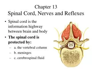
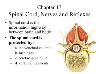
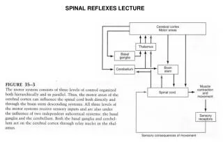
Integrating center for the reflex activity between afferent input and efferent output is located in one or more spinal cord segments • The brain can facilitate or inhibit them • Examples: • Stretch reflex Spinal reflexes
Two muscle receptors are important for proprioceptive inputs: • Muscle spindles (monitor changes in muscle length) • Golgi tendon organs ( monitor changes in muscle tension) Stretch reflex
Distributed throughout skeletal muscle fibers • Each spindle consists of 3-10 specialized muscle fibers enclosed in a connective tissue capsule (intrafusal fibers) • Each intrafusal fiber has • Noncontractile central portion • Contractile ends Muscle spindles Muscle spindle skeletal muscle fibers
Muscle spindles • Each spindle has: • Afferent nerve supply • sensory nerve endings detect change in muscle length and speed • Efferent nerve supply • motor, gamma neurons Skeletal muscle fibers are supplied via α motor neurons
Patellar tendon reflex (a stretch reflex) 2 Sensory from spindles Tapping stretches the muscle spindles in the quadriceps femoris muscle 1 3 To skeletal muscle fibers
In the tendons of the muscle • Respond to changes in the muscle’s tension • Increased firing with increased muscle tension • Its firing leads to inhibition of α motorneuron and thus relaxation of skeletal muscle Golgi tendon organs
1 Increased tension increases GTO firing 2 Contraction increases muscle tension 3 4 Inhibitory motorneuron 5 Muscle relaxes
Opposite of those elicited by muscle spindle reflexes • Golgi tendon organs help ensure smooth onset and termination of muscle contraction • Particularly important in activities involving rapid switching between flexion and extension such as in running Golgi tendon reflex